
Help Bill! Bioprinting Skin, Muscle and Bone
Students operate mock 3D bioprinters in order to print tissue constructs of bone, muscle and skin for a fictitious trauma patient, Bill. The model bioprinters are made from ordinary materials— cardboard, dowels, wood, spools, duct tape, zip ties and glue (constructed by the teacher or the students)—and use squeeze bags of icing to lay down tissue layers. Student groups apply what they learned about biological tissue composition and tissue engineering in the associated lesson to design and fabricate model replacement tissues. They tangibly learn about the technical aspects and challenges of 3D bioprinting technology, as well as great detail about the complex cellular composition of tissues. At activity end, teams present their prototype designs to the class.
3D bioprinting is a novel engineer-created biomedical technology with the potential to revolutionize the field of tissue engineering and ultimately, organ transplantation. Currently, biomedical engineers and researchers use this technology to print and experiment with simpler tissues, with the aim to be able to print functional, complex organs in the future. By participating in this activity, students experience similar challenges as technology researchers and inventors, including setting workable printing parameters and accounting for blood vessels.
After this activity, students should be able to:
- Operate a model/mock 3D bioprinter to print a model graft of skin, muscle or bone.
- Explain what 3D bioprinting is and describe the importance of this technology.
- Identify the different types of cells and structural components necessary to print a model graft of skin, muscle or bone.
- Evaluate the strengths and weaknesses of the prototype model grafts and of the bioprinting process.
- CCSS.Math.Content.HSG-GMD.B.4 Identify the shapes of two-dimensional cross-sections of three-dimensional objects, and identify three-dimensional objects generated by rotations of two-dimensional objects.
Grades 9-12
Do you agree with this alignment? - CCSS.Math.Content.HSG-MG.A.1 Use geometric shapes, their measures, and their properties to describe objects (e.g., modeling a tree trunk or a human torso as a cylinder).
Grades 9-12
Do you agree with this alignment? - CCSS.Math.Content.HSG-MG.A.3 Apply geometric methods to solve design problems (e.g., designing an object or structure to satisfy physical constraints or minimize cost; working with typographic grid systems based on ratios).
Grades 9-12
Do you agree with this alignment?
- Design problems are seldom presented in a clearly defined form.
Grades 9-12
Do you agree with this alignment? - STEL-7AA Illustrate principles, elements, and factors of design.
Grades 9-12
Do you agree with this alignment?
- DCI.LS1.A.9-12.6 Systems of specialized cells within organisms help them perform the essential functions of life.
Grades 9-12
Do you agree with this alignment? - DCI.LS1.A.9-12.8 Multicellular organisms have a hierarchical structural organization, in which any one system is made up of numerous parts and is itself a component of the next level.
Grades 9-12
Do you agree with this alignment?
- The student will investigate and understand relationships between cell structure and function. Key concepts include
Grades 9-12
Do you agree with this alignment?
Each group needs:
- 1 mock bioprinter, constructed in advance by the teacher OR during the class by students, using components available at craft stores, such as Michaels; required components:
- wooden platform, 12 x 12 inches (30 x 30 cm)
- 4 wooden spools, each 2.75-inches tall (7 cm)
- 8 wooden rods/dowels, 12-inches long x 3/8-inch diameter (30 x 1 cm)
- 4 pieces of duct tape, each 1 x 2 inches (2.54 x 5 cm)
- 2 balsa wood or basswood arms, each 10 x 3 inches (25 x 7.6 cm)
- 3+ cardboard cutouts to use as “stages” and to raise/lower the printing platform
- 12 zip ties
- wood glue
- 1 background information sheet on skin, bone OR muscle, from Injury Area Information Sheets (PDF)
- 3D Bioprinting Parameters Worksheet (PDF)
- Post-Activity Quiz (PDF), one per student
To share with the entire class:
- ~5 colors of icing, in many icing bags with tips, which represent different types of cells and extracellular matrix
- ability to show the class a few short videos; see URLs in the Additional Multimedia Support section
- other supplies and tools that students request for their designs or process, such as additional pieces of cardboard, tape, rulers, etc.
A basic understanding of cell theory; basic information about 3D bioprinting as provided in the associated lesson.
(To kick off the activity, show the class a 51-second video of trauma patient, Bill, at https://www.youtube.com/watch?v=XyZozl9poto.)
Imagine that the year is 2050 and Bill has been injured in an automobile accident. A section of his bone, muscle and skin were damaged beyond repair and need to be totally replaced by 3D bioprinting. You have been hired as biomedical engineers to help print bone, muscle and skin grafts for Bill.
Biomedical engineers must consider the cellular composition, extracellular matrix, vascularization, and shape of tissues in order to ensure that the bioprinted replacement tissue provides the desired structure and function. Your group will be assigned to print a section of either bone, skin or muscle for Bill using 3D bioprinting. These grafts, if printed correctly, increase the likelihood that Bill will heal correctly and be restored to full health more quickly.
During the accident, Bill lost approximately one cubic inch of skin on his left arm, covering four square inches. Human skin provides multiple vital physiological functions, including body temperature regulation, protection from the environment, and water retention. Skin has three main layers: epidermis, dermis and hypodermis (see Figure 1).
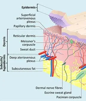 Figure 1. A diagram shows the skin tissue layers. Bill has injured skin.Copyright 2013 Anatomy Box, Creative Commons Attribution Share License http://www.anatomybox.com/chapters/skin/ http://www.anatomybox.com/wp-content/uploads/2012/08/hair-follicles-thick-thin-skin.jpg
During the accident, an approximately one-inch section of Bill’s femur was completely shattered beyond repair; the diameter of the femur is approximately one inch. The femur is the long bone that runs down the upper leg and is crucial for walking (see Figure 2). We believe that we can produce a new section of bone for Bill using a 3D bioprinter. By introducing it between the two healthier pieces, then putting the leg in a cast for about a year, his body will connect the three bone pieces and make it whole again. The femur is a “hard bone,” which takes on a lot of the stress from bearing Bill’s weight. Hard bone has three main layers: periosteum, cortical/compact bone, and medullary cavity.
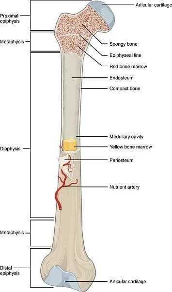 Figure 2. A drawing of a long bone and its underlying tissue. Bill has a broken femoral shaft.Copyright 2016 Carl Fredrick, OpenStax Anatomy and Physiology, Wikimedia Commons CC BY-SA 4.0 https://commons.wikimedia.org/wiki/File:603_Anatomy_of_a_Long_Bone.jpg https://creativecommons.org/licenses/by/4.0/deed.en
Bill also lost three cubic inches of his rectus femoris, one of the large “skeletal muscles” that controls his femur (the bone in his upper leg; see Figure 3). The rectus femoris is part of the quadricep group and is crucial for walking and running. By printing a new section of muscle, we can restore Bill’s leg to its previous strength. Studies have shown that nerves “innervate” (connect to and control) replaced muscle tissue after implantation. Skeletal muscle is made of many tube-shaped skeletal muscle fibers, all stacked in neat rows.
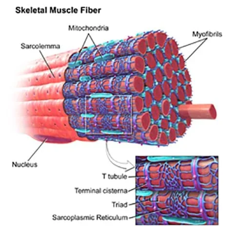 Figure 3. A drawing of a muscle. Bill has a ripped femoral muscle.Copyright 2014 WikiJournal of Medicine Gallery of Blausen Medical CC BY-NC-ND-4.0 http://teachmeanatomy.info/the-basics/ultrastructure/histology-muscle/ https://creativecommons.org/licenses/by-nc-nd/4.0/
Bill is counting on your team of biomedical engineers to bioprint bone, muscle and skin grafts to help him recover from the accident. Let’s get started!
Activity Overview
In this activity, students act as if they are biomedical engineers printing replacement skin, muscle or bone grafts for a fictitious emergency room patient. The overarching steps of the project are:
- Groups review their tissue assignments (skin, bone, muscle). (~5 minutes)
- They learn to use their mock bioprinters. (~2 minutes)
- They make engineering sketches of their plans and get teacher approval. (~10 minutes)
- Teams get biomaterials and print! (~20 minutes)
- They present to the class their final prototype designs and limitations. (~2 minutes per group)
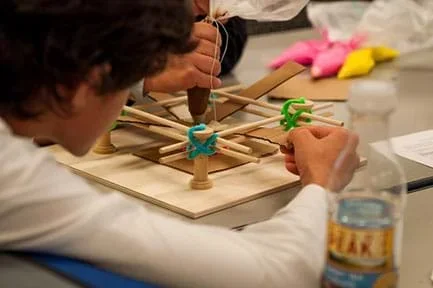 Teenagers operate a mock-3D bioprinter to print skin for Bill.Copyright 2017 Hunter Sheldon, Department of Biomedical Engineering, University of Virginia
Before the Activity
- Assemble the model/mock bioprinters using the seven-minute Mock-3D Bioprinter Assembly Instructions video at https://www.youtube.com/watch?v=j_De-LaTrPo. Make enough bioprinters so that each group of four students has one. Alternatively, have students construct their own bioprinters.
- Fill the icing bags with different colored icing, representing different cell types. See below for suggestions for five different colors, what they represent, and the distribution of icing bags per group/tissue type. Label the bags so that students can easily select the correct biomaterials for their assigned tissue types. Create enough icing bags so all teams can be working simultaneously.
- Make copies of the 3D Bioprinting Parameters Worksheet (PDF) (one per group), Post-Activity Quiz (PDF) (one per student), and Injury Area Information Sheets (PDF) (includes skin, bone, and muscle sheets; make enough for one tissue type per group).
- Think about how you will divide the class into groups of four students each. To accommodate differing class sizes, groups of three to five students also work. We strongly recommend not creating groups that are larger than five students or smaller than three students.
- Ideally, give each group an individual space to work and write.
- Place a mock-3D bioprinter at each group station. Include at least three cardboard printing “stages,” which gives teams enough to start over if necessary, stack for increased vertical height, etc.
- If the lesson and activity are conducted in one class session, prepare the activity setup before starting the lesson.
- If the lesson and activity are conducted in two separate class periods, spend some time reviewing the lesson material before beginning the activity.
Preparation of Icing Colors
The colors listed below are based on the icing colors that are commonly found at grocery stores and how closely they represent the natural material color. Feel free to substitute or change as desired.
Bone: Each group needs one of each of the following icing bags:
- Cortical bone: vanilla/white
- Periosteum: buttercream
- Vasculature: pink
- Yellow marrow: yellow
Skin: Each group needs one of each of the following icing bags:
- Hypodermis: vanilla/white
- Epidermis: buttercream
- Dermis: yellow
- Vasculature: pink
- Hair/sweat glands: brown
Muscle: Each group needs one of each of the following icing bags:
- Myocytes: brown
- Support material: buttercream
- Vasculature: pink
With the Students
- Divide the class into groups of four students each.
- Intro Scenario: Introduce the engineering project through the PowerPoint® slides in the associated lesson and/or by presenting the Introduction/Motivation content, both of which kick off the activity with a video about fictitious trauma patient, Bill.
- Review: As makes sense for your class, but especially if the associated lesson was taught a day or more before the activity is conducted, spend some time reviewing the key concepts and the hypothetical design challenge scenario using the pre-activity questions provided in the Assessment section.
- Skill Prep: Show students how to operate the 3D bioprinter by showing them the 50-second Mock-3D Printer Bioprinter Instructions for Use video at https://www.youtube.com/watch?v=iNKIGC7OooI.
- Design Assignment: Assign each group either skin, muscle or bone. Hand out to each team a worksheet and the appropriate injury area background sheet.
- Give students some time to read over both handouts, become familiar with their specific tissues, and talk in their teams about what biomaterials they need to construct their specific tissues.
- Then have a member from each group collect the necessary biomaterials (icing bags) that his/her group needs for the activity and bring them to the group’s station.
- Design/Planning: Give the groups ~10 minutes to plan and sketch their designs. If available, have students sketch their design ideas in their design notebooks. The planning effort includes drawing a labeled engineering sketch of the design that is complete with identified biomaterials, measurements, cross-sections, etc. For more information on engineering sketches, refer to the Detail Drawings: Communicating with Engineers lesson.
- As students work on their designs, move amongst the teams to make sure they work collaboratively and stay on task. Ask them the Investigating Questions.
- Have each group show you its design before permitting them to proceed.
- Make sure each team is using the correct biomaterials for the assigned tissue type and have a general plan of action for how to print the tissue. The important thing is to make sure they are not going into the printing process blind!
- Make sure they go through the worksheet, which prompts them to consider some bioprinting parameters such as printing pressure, speed, nozzle, path, and icing viscosity.
- Once a design is approved, give the team a bioprinter.
- Fabrication/Iteration: Give students time to use their bioprinters to print various designs onto their cardboard stages. Make sure students are filling out the worksheet during the printing process. Tip: Let the students bioprint! Give them plenty of time and space to try new things and make mistakes.
- As they work, have them assign one team member to take notes on the experience. Ask: What about the printing process is difficult? What is easy? What problems did you encounter? What solutions did you figure out? These notes will be important for their final class presentations.
- If a group finishes early, encourage/challenge them to make a different design/prototype, changing the printing orientation or other factors.
- If a group is taking too long or not making progress, encourage them not to be afraid to make mistakes. Remind them that we learn from mistakes and experimentation, and that continuous iteration—repeated adjustments and redesigns to get closer to the desired result—is part of the engineering design process.
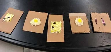 Example final designs of mock-3D printed tissues from five student groups. From left to right: skin, bone, skin, bone, muscle.Copyright 2016 Angela Sickels. School of Engineering and Applied Science. University of Virginia
- With ~15 minutes left in class, end the printing and have teams each give a quick oral presentation, as described in the Assessment section.
- Administer the post-activity quiz, as described in the Assessment section.
- Clean-up:
- Dispose of any cardboard printing stages that are covered in mock biomaterial.
- Dispose of any bags still filled with biomaterials, unless you plan on doing the activity again within a week.
- Rinse the icing bags tips so they are ready to be used again.
- Store the mock bioprinters until next use.
- 3d bioprinting
- A way of printing tissues, layer by layer. This printed tissue contains two parts: the cells and the unique mixture of fibers that composes its structure and shape.
- bio-ink
- Cells in a medium that makes them suitable for printing in an inkjet bioprinter. This liquid substance solidifies after printing.
- biomaterial
- A substance that is engineered to interact with the components of living biological systems for therapeutic or diagnostic medical purposes.
- bone graft
- A surgical procedure that replaces bone tissue, especially in cases of trauma-damaged bones.
- extracellular matrix
- The non-cellular matrix of fibrous biomaterials. This matrix gives body tissues support as well as various mechanical properties. Abbreviated as ECM.
- iteration
- Doing something again. Common in the engineering design process by making continuous and repeated adjustments and redesigns to get closer to the desired result.
- muscle graft
- A piece of muscle, typically used in complex reconstruction surgeries. Also referred to as a “flap technique.”
- skin graft
- Transplanted skin tissue.
- transplant rejection
- When the body attacks transplanted tissue because the transplant is perceived as foreign by the immune system. Also called immune rejection.
- vascularization
- The formation of blood vessels, either naturally or synthetically.
- viscosity
- A measure of a fluid’s resistance to flow due to internal friction. A measure of a fluid’s “thickness.”
Pre-Activity Assessment
Review: Review the key concepts learned from the associated lesson, ask students the following questions. Find out what they retained about these concepts and fill in their knowledge gaps as necessary.
- What is 3D bioprinting?
- Who is Bill? What happened to Bill?
- What type of bioprinter will we be using? (Answer: Extrusion)
- What are the five design steps of tissue engineering that we will use? (Answer: 1] identify function being replaced, 2] determine cell types, 3] determine biomaterials types, 4] determine construction method, and 5] construction/fabrication.)
- What are some limitations of bioprinting? What makes it challenging? (Answer: Addressing the issues of vascularization, immune rejection, and biocompatibility; hard to print functional tissues at such a small scale; other possible limitations such as printing speed, multiple layering, material waste, cell viability, resolution, viscosity, density.)
- What is the point of including biomaterials in our designs? What purpose do they serve in our printed tissues? (Answer: Ideal biomaterials mimic and function like real human tissues, are biocompatible, do not degrade into toxic materials in the body, and are less likely to be rejected by human bodies.)
Activity Embedded Assessment
In-Class Design Presentations: Give each group two minutes to give an oral presentation of its tissue construct and explain its reasoning, as well describe a few design process steps they went through to create the tissue. Discuss successes and difficulties, connecting the design process they just experienced to the real-world 3D bioprinting process.
1. How does your design compare to the structure of real biological tissue?
2. What was difficult about the printing process?
3. How could you have improved your design?
4. What would you change about the printer to make the printing process easier or more efficient?
Post-Activity Assessment
Post-Quiz: After the activity is completed, administer the four-question Post-Activity Quiz (PDF) in class if time permits, or as homework that night, so the activity is still fresh in students’ minds. The quiz asks them to reflect on their 3D bioprinting experiences—describe what tissue they printed, which tissue features were hardest to print, what was most challenging about 3D bioprinting, and how they might improve upon their designs. It also includes an optional activity extension—to choose another tissue that could be 3D bioprinted, research it, and write up a brief printing plan.
Remind the “junior biomedical engineers” not to eat their materials.
Especially after one use, the icing bag tips may get clogged. If not unclogged, students may have a hard time printing during the activity and may even pop an icing bag. If you plan to repeat the activity and reuse the tips with new icing bags, make sure to clean them by running them under hot water and using a toothpick to poke out any dried icing.
Instead of hand-drawn sketches, have students use Google Sketchup or other CAD software to design their tissue constructs.
If time permits, have groups print all three tissue types and reflect on the feasibility and difficult of printing each one.
For lower grades, do not assign muscle as a tissue to 3D print because it is the most difficult to design.
For more advanced students, have them construct the mock 3D bioprinters.
Use these YouTube videos to prepare for and conduct the activity:
1. Help Bill! Bioengineering Challenge (1:05 seconds): https://www.youtube.com/watch?v=XyZozl9poto
2. Mock-3D Bioprinter Assembly Instructions (7:23 minutes):https://www.youtube.com/watch?v=j_De-LaTrPo
3. Mock-3D Bioprinter Instructions for Use (1:04 seconds): https://www.youtube.com/watch?v=iNKIGC7OooI
Buntic, Rudolf. Vascularized Muscle Flaps. Microsurgeon.org. Accessed February 17, 2017. https://www.microsurgeon.org/muscleflaps
Frantz, Christian, Stewart, Kathleen M. & Weaver, Valerie M. Published 2010. “The extracellular matrix at a glance,” Journal of Cell Science. Accessed February 17, 2017. Vol. 123, No. 24, pp. 4195-4200. https://doi.org/10.1242/jcs.023820
Murphy, Sean V. & Atala, Anthony. Published August 5, 2014. “3D bioprinting of tissues and organs.” (2014) Nature Biotechnology. Vol. 32, pp. 773–85. DOI: doi:10.1038/nbt.2958 http://www.nature.com/nbt/journal/v32/n8/full/nbt.2958.html
Skin grafting. Last updated January 4, 2017. Wikipedia, the free encyclopedia. Accessed February 17, 2017. https://en.wikipedia.org/w/index.php?title=Skin_grafting&oldid=758352543
- Intro to 3D Bioprinting: Design, Applications and Limitations
- Help Bill! Bioprinting Skin, Muscle and Bone
Contributors
Nicholas Asby; Angela Sickels; Hunter Sheldon; Ryan Tasker-Benson; Timothy Allen; Shayn M. Peirce; A. L. Peirce Starling
Supporting Program
Department of Biomedical Engineering, School of Engineering and Applied Sciences, University of Virginia
Acknowledgements
This digital library curriculum—an engineering tool kit (ETK) for high school students—was developed under a grant from The Jefferson Trust. Special thanks to Michaela Rikard and Vi Tran of the UVa Department of Biomedical Engineering for their assistance throughout the project.
Copyright
2017 by Regents of the University of Colorado; original © 2016 University of Virginia