
Engineering Nature: DNA Visualization and Manipulation
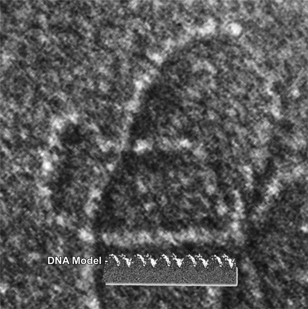 Electron micrograph of a DNA sample.Copyright 2012 University of California Los Angeles http://www.biolchem.ucla.edu/Resources_Electron_Microscopy_Laboratory_Images.htm
This unit presents technologies used to visualize the complex structures of biomolecules such as DNA and proteins, as well as DNA fingerprinting by gel electrophoresis, a DNA analysis technique used in forensic studies.
In the first lesson, "Imaging DNA Structure," students are introduced to imaging methods in current use and the challenges faced by biologists and biomedical engineers when trying to understand biomolecule structures. In the associated activity, students research specific imaging technologies with applications in biology and present their findings.
In the second lesson, "Restriction Enzymes and DNA Fingerprinting," students learn about electrophoresis applications for DNA fingerprinting. The associated hands-on activity illustrates the concept of electrophoresis and DNA fingerprinting.
Visualization of small structures such as the molecular structures of complex proteins and genetic material (DNA) is possible due to engineering discoveries and breakthroughs in physics at small scales. Imaging technologies such as x-ray and scanning electron microscopy—used by scientists and engineers to image microscopic structures—are also used by biomedical engineers and biologists to study biomolecules, cells and tissue samples. Microfluidics concepts and devices used to study colloidal particle flow are also employed by biologists to study and filter biomolecules. Gel electrophoresis is one example engineering application that is used by biologists to compare fragments of DNA samples.
- Technological progress promotes the advancement of science and mathematics.
Grades 9-12
Do you agree with this alignment? - The sciences of biochemistry and molecular biology have made it possible to manipulate the genetic information found in living creatures.
Grades 9-12
Do you agree with this alignment?
- know that hypotheses are tentative and testable statements that must be capable of being supported or not supported by observational evidence. Hypotheses of durable explanatory power which have been tested over a wide variety of conditions are incorporated into theories;
Grades 9-11
Do you agree with this alignment? - know scientific theories are based on natural and physical phenomena and are capable of being tested by multiple independent researchers. Unlike hypotheses, scientific theories are well-established and highly-reliable explanations, but they may be subject to change as new areas of science and new technologies are developed;
Grades 9-11
Do you agree with this alignment? - distinguish between scientific hypotheses and scientific theories;
Grades 9-11
Do you agree with this alignment? - plan and implement descriptive, comparative, and experimental investigations, including asking questions, formulating testable hypotheses, and selecting equipment and technology;
Grades 9-11
Do you agree with this alignment? - analyze the levels of organization in biological systems and relate the levels to each other and to the whole system.
Grades 9-11
Do you agree with this alignment?
- Day 1: Imaging DNA Structure lesson
- Day 2: Inside the DNA activity
- Day 3-4: Restriction Enzymes and DNA Fingerprinting lesson
- Day 5: DNA Forensics and Color Pigments activity
- Engineering Nature: DNA Visualization and Manipulation
Contributors
Mircea Ionescu; Myla Van Duyn
Supporting Program
National Science Foundation GK-12 and Research Experience for Teachers (RET) Programs, University of Houston
Acknowledgements
This digital library content was developed by the University of Houston's College of Engineering under National Science Foundation GK-12 grant number DGE-0840889. However, these contents do not necessarily represent the policies of the NSF and you should not assume endorsement by the federal government.
Copyright
2013 by Regents of the University of Colorado; original © 2012 University of Houston