
Using Stress and Strain to Detect Cancer!
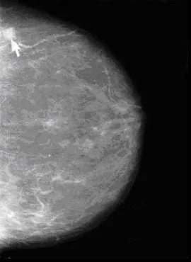 A mammogram shows a tumor.Copyright National Institutes of Health (public domain image)
The unit's design uses a contextually based "Challenge" followed by a sequence of instruction in which students first offer initial predictions ("Generate Ideas") and then gather information from multiple sources ("Multiple Perspectives"). This is followed by "Research and Revise" as students integrate and extend their knowledge through a variety of learning activities. The cycle concludes with formative ("Test Your Mettle") and summative ("Go Public") assessments that lead the student towards answering the Challenge question. See the unit overview below for the progression of the legacy cycle through the unit. Research and ideas behind this way of learning may be found in How People Learn, (Bransford, Brown & Cocking, National Academy Press, 2000); see the entire text at http://www.nap.edu/html/howpeople1/.
The legacy cycle has similarities to the engineering design process; they both involve identifying needs existing in society, combining science and math to develop solutions, and applying the research conclusions to design clear conceived solutions to the challenges. Though the engineering design process and the legacy cycle depend on a correct and accurate solution, each focuses particularly on how the solution is devised and presented. See an overview of the engineering design process at http://en.wikipedia.org/wiki/Engineering_design_process.
In lesson 1, students are presented with the following Grand Challenge: "Breast cancer is the second-leading cause of cancer death among women (Papas, 253) and the American Cancer Society has indicated that mammography is the best early-detection tool available. Despite the fact that mammograms are the most effective early-detector of breast cancer, many women choose not to have them. Of all American women at or over the age of 40, only 54.9% have had a mammogram within the past year (ACS, 15). One reason women attribute skipping their annual mammogram is pain with 90% report experiencing discomfort (Papas, 254). Is there a way to detect the presence of tumors that isn't as painful as mammography but more reliable and quantifiable than a breast-self exam and clinical breast-exams?"
Students begin by Generating Ideas in lesson 1's associated activity, answering questions such as, "What are your initial thoughts about how this question can be answered?" and "What do you know about breast cancer and tumor detection already?" Students then enter the Multiple Perspectives phase of the legacy cycle as they read an expert interview from a professor of biomedical imaging. To extend the Multiple Perspectives phase, it is suggested that a local specialist make a guest presentation on medical imaging to the class.
In lesson 2, students enter the Research and Revise phase focusing on the concepts of Hooke's law and stress-strain relationships. The lesson includes a problem set to complete individually or as a class. The practice problems give students experience manipulating the new equations. Students extend their understanding of these concepts in the associated activity. In the first portion of the activity, students explore Hooke's law by experimentally solving for an unknown spring constant. In the second portion of the activity, students apply this understanding to study breast tissue with respect to cancer detection. After completing the lesson and the associated activities, students enter the Test Your Mettle phase by completing a quiz on stress, strain and Hooke's law.
Finally, lesson 3 and its associated activity constitute the Go Public phase of the legacy cycle. First, in the associated activity, students create a graph in Excel® depicting the location of a cancerous tumor amidst healthy breast tissue. This in-class assignment constitutes one-half of the grade while the other half results from a take-home assignment. In the take-home assignment, students are create informative brochures presenting their innovative, painless form of breast cancer detection to patients and physicians.
Today, medical imaging is generally accepted as a safe, noninvasive means of generating internal images of the body. More than 100 years ago, such beneficial uses of harmful radiation would have been dismissed as outrageous and impossible. Decades of work by doctors, radiologists, scientists and engineers has provided safer use of x-rays for imaging of the skeletal system and some soft tissue, computer axial tomography, CT scans, to obtain three dimensional x-ray based imaging, magnetic resonance imaging, MRI, for depicting high contrast in soft tissues, and ultrasound imaging using high-frequency sound waves to depict moving structures and tissues in real time. Unfortunately, challenges remain in medical imaging today; one of which is presented in this unit. With nearly 45% of women avoiding annual mammograms due to pain and discomfort, students are challenged to think like engineers to develop a painless means of breast cancer detection that is reliable, cost effective and noninvasive.
- CCSS.Math.Content.HSF-IF.B.6 Calculate and interpret the average rate of change of a function (presented symbolically or as a table) over a specified interval. Estimate the rate of change from a graph.
Grades 9-12
Do you agree with this alignment?
- HS-ETS1-1 Analyze a major global challenge to specify qualitative and quantitative criteria and constraints for solutions that account for societal needs and wants.
Grades 9-12
This resource focuses on the following Three Dimensional Learning aspects of NGSS:
Science & Engineering Practices- Analyze complex real-world problems by specifying criteria and constraints for successful solutions.Do you agree with this alignment?
Disciplinary Core Ideas- Criteria and constraints also include satisfying any requirements set by society, such as taking issues of risk mitigation into account, and they should be quantified to the extent possible and stated in such a way that one can tell if a given design meets them.Do you agree with this alignment?
- Humanity faces major global challenges today, such as the need for supplies of clean water and food or for energy sources that minimize pollution, which can be addressed through engineering. These global challenges also may have manifestations in local communities.Do you agree with this alignment?
Crosscutting Concepts- New technologies can have deep impacts on society and the environment, including some that were not anticipated. Analysis of costs and benefits is a critical aspect of decisions about technology.Do you agree with this alignment?
Do you agree with this alignment? - Analyze complex real-world problems by specifying criteria and constraints for successful solutions.
- HS-ETS1-2 Design a solution to a complex real-world problem by breaking it down into smaller, more manageable problems that can be solved through engineering.
Grades 9-12
This resource focuses on the following Three Dimensional Learning aspects of NGSS:
Science & Engineering Practices- Design a solution to a complex real-world problem, based on scientific knowledge, student-generated sources of evidence, prioritized criteria, and tradeoff considerations.Do you agree with this alignment?
Disciplinary Core Ideas- Criteria may need to be broken down into simpler ones that can be approached systematically, and decisions about the priority of certain criteria over others (trade-offs) may be needed.Do you agree with this alignment?
Do you agree with this alignment? - Design a solution to a complex real-world problem, based on scientific knowledge, student-generated sources of evidence, prioritized criteria, and tradeoff considerations.
- Day 1: Your Biomedical Challenge: Painlessly Detecting Disease lesson
- Day 2: Learning Imaging Techniques! activity
- Day 3: Stress, Strain and Hooke's Law lesson
- Day 4-5: Applying Hooke's Law to Cancer Detection activity
- Day 6: Making Brochures: Presenting Painless Cancer Detection! lesson and You Be the Radiologist! Strain Graphs That Reveal Tumors activity
- Using Stress and Strain to Detect Cancer!
Contributors
Meghan Murphy; Luke Diamond
Supporting Program
VU Bioengineering RET Program, School of Engineering, Vanderbilt University
Acknowledgements
The contents of this digital library curriculum were developed under National Science Foundation RET grant nos. 0338092 and 0742871. However, these contents do not necessarily represent the policies of the NSF, and you should not assume endorsement by the federal government.
Copyright
2013 by Regents of the University of Colorado; original © 2007 Vanderbilt University