
Exploring Bone Mineral Density, X-Rays and Biomedical Imaging
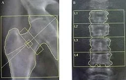 A dual-energy x-ray absorptiometry (DEXA) assessing bone mineral density.Copyright 2010 Caroline Lebreton and Raymond Poincaré Chu, Wikimedia Commons http://commons.wikimedia.org/wiki/File:Morbus_Fabry_DXA_01.jpg
Student teams explore two websites to gather information on bone mineral density and how it is measured. They also learn about x-rays in general, how they work and their different uses, along with other imaging modalities. They answer guiding questions as they explore the websites and take a short quiz afterwards to test the knowledge they gained while reading the articles.
The topics of bone mineral density and x-rays are both important to biomedical engineers and are at the forefront of many areas of current research. In this initial gathering of information stage, students begin to build their knowledge base in order to come closer to answering the unit challenge question. Having a good background on how a system works is necessary to make critical decisions on its products, which in this case is an x-ray image.
After this activity, students should be able to:
- Explain bone mineral density, its uses and tests.
- Explain how an x-ray works.
- Telemedicine reflects the convergence of technological advances in a number of fields, including medicine, telecommunications, virtual presence, computer engineering, informatics, artificial intelligence, robotics, materials science, and perceptual psychology.
Grades 9-12
Do you agree with this alignment? - STEL-3F Apply a product, system, or process developed for one setting to another setting.
Grades 6-8
Do you agree with this alignment?
- Scientific Research
Grades 9-12
Do you agree with this alignment?
Each group needs:
- Copy of question sheet.
- Access to computer or printed copy of both articles.
Now that we have brainstormed a little about the challenge question, you have told me that we all need to learn more about bone mineral density and how it is measured with x-rays or otherwise. So today you are going to read a few articles and answer some questions as you go to learn more about BMD and x-rays. You might need to finish as homework. Tomorrow we will have a short quiz covering the material from the articles and from your questions you will answer.
Before the Activity
- Make copies of the question sheet, one per student.
- Bookmark the websites on the computers the students will use, or make paper copies of the articles to hand out to each student.
With the Students
Ask students to navigate through the website https://www.webmd.com/osteoporosis/guide/bone-mineral-density#1 to gather information regarding what BMD is and why it is important. Using the website https://science.howstuffworks.com/x-ray.htm, direct students through a brief overview of the history and mechanics of x-rays. Be sure to point out to students the other uses of x-rays. Students should answer a series of questions as they are reading. Ask the students to turn in these questions and take a short quiz over what they have read.
- bmd
- Acronym for bone mineral density. The density of minerals (such as calcium) in bones.
- osteopenia
- The natural thinning of bones over time.
- dual-energy x-ray absortiometry
- A method used to calculate bone mineral density that relies on two different X-ray beams passing through an object so that the materials inside the object can be distinguished (such as bone and soft tissue). Abbreviated as DEXA or DXA.
- attenuation
- The reduction in intensity of an x-ray beam as it passes through a material.
- attenuation coefficient
- The measure of the reduction of the intensity of an x-ray beam that depends on the material of the object and the intensity of the x-ray beam.
Post-Activity Assessment: Administer the BMD Reading Quiz. Review students' answers to gauge their depth of comprehension.
After students have completed the article worksheet, ask them again for their thoughts on the challenge question. Ask if they have any ideas to add now that they have done some research.
- Bone Mineral Density and Logarithms
- Bone Density Challenge Introduction
- Exploring Bone Mineral Density, X-Rays and Biomedical Imaging
- Bone Density Math and Logarithm Introduction
- Common and Natural Logarithms and Solving Equations
- Bone Mineral Density Math and Beer's Law
- Bone Density Challenge Introduction
Contributors
Kristyn Shaffer; Megan Johnston
Supporting Program
VU Bioengineering RET Program, School of Engineering, Vanderbilt University
Acknowledgements
The contents of this digital library curriculum were developed under National Science Foundation RET grant nos. 0338092 and 0742871. However, these contents do not necessarily represent the policies of the NSF, and you should not assume endorsement by the federal government.
Copyright
2013 by Regents of the University of Colorado; original © 2006 Vanderbilt University