
Bone Density Challenge Introduction
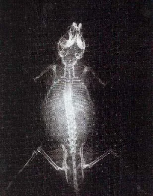 An x-ray iimage of a mouse, obtained via cabinet x-ray system.Copyright Dr. Edwin Donnelly, Vanderbilt University Medical Center
Students are introduced to the challenge question, which revolves around proving that a cabinet x-ray system can produce bone mineral density images. Students work independently to generate ideas from the questions provided, then share with partners and then with the class as part of the Multiple Perspectives phase of this unit. Then, as part of the associated activity, students explore multiple websites to gather information about bone mineral density and answer worksheet questions, followed by a quiz on the material covered in the articles.
Medical imaging is a large area of study for biomedical engineering. As students explore one of the many modalities of medical imaging, x-rays, they begin the think like engineers when they gather information in their research, just as engineers research before proceeding to imagine and make decisions as part of the design process. In addition, the challenge question forces students make a critical decision based on criteria that they learn throughout the unit.
After this lesson, students should be able to:
- Explain the importance and uses of bone mineral density.
- Explain how x-rays work.
- Explain how logarithms connect to bone mineral density tests.
- Identify the design problem to solve and decide whether or not to address it.
Grades 9-12
Do you agree with this alignment? - STEL-7Y Optimize a design by addressing desired qualities within criteria and constraints.
Grades 9-12
Do you agree with this alignment?
- Biology I
Grades 9-12
Do you agree with this alignment? - Scientific Research
Grades 9-12
Do you agree with this alignment?
Introduce the challenge question and hand out the worksheet to help students in generating ideas. The worksheet lists the following questions for guidance:
- What are your initial ideas about how these questions can be answered?
- What background is needed?
- What do you know about x-rays already?
- What do you know about bone mineral density already?
In the class setting, have students share ideas from their journals. Record their ideas on the classroom board. Work with the class to set up main categories of necessary knowledge. If possible, organize the topics into the following three categories: focus on bone density, imaging, the mathematics involved in calculating a BMD image.
Then move on to the associated activity, Exploring Bone Mineral Density, X-Rays and Biomedical Imaging to have students explore some websites about bone mineral density.
The teacher should be familiar with how a BMD image is created using a small-specimen x-ray system. Refer to the following websites for a brief overview to prepare for this lesson and the associated activity.
Your engineering challenge question: You work for a company that manufactures cabinet x-ray systems used in imaging small specimens. You want to sell more systems, so you need to find a way to make your current system more marketable by showing that your system is capable of performing multiple functions. One function you want to investigate is if your current system is capable of producing bone mineral density (BMD) images. If it is, you believe that you can increase your sales dramatically. You hire a team of biomedical engineers to research your product's applications to BMD. After many hours of research, the team has presented the following image to you. Is this an acceptable image? How do you know?
Grade students on their written journal brainstorming and class participation in the class brainstorming.
Contributors
Kristyn Shaffer; Megan Johnston
Supporting Program
VU Bioenegineering RET Program, School of Engineering, Vanderbilt University
Acknowledgements
The contents of this digital library curriculum were developed under National Science Foundation RET grant nos. 0338092 and 0742871. However, these contents do not necessarily represent the policies of the NSF, and you should not assume endorsement by the federal government.
Copyright
2013 by Regents of the University of Colorado; original © 2006 Vanderbilt University