Quick Look
Grade Level: 7 (6-8)
Time Required: 2 hours
(90 minutes to develop and test, 30 minutes to review results)
Expendable Cost/Group: US $15.00
Group Size: 3
Activity Dependency: None
Subject Areas: Biology, Chemistry, Life Science, Number and Operations, Physics
NGSS Performance Expectations:

| MS-ETS1-4 |
| MS-LS1-1 |
Summary
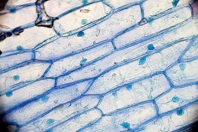
In this design analysis activity, students make sense of technologies used to help visualize cell functions down to the nanometer scale such as con-focal microscopes, lasers, and customized fluorescent dyes. Students design and test new ideas to image plant cells using common household items and improve upon the process with testing. Using the iodine solution process as a control, student teams plan how to image the cells, evaluate the results and make recommendations to improve the imaging process.Engineering Connection
Adding a black light and fluorescent dyes images better to find is a way to better image onion cells. This is a basic version of using con-focal microscopy to image cells. The objectives in this activity are in line with engineering design process. The steps include identifying a need; developing possible solutions; plan a prototype; test and evaluate; and optimize the solution. The key to this activity is to have students learn on their own and develop an understanding of the engineering process and come up with their own conclusions of the activity. Though some solutions may be better, it’s up to the student to determine the possible solutions.
Learning Objectives
After this activity, students should be able to:
- Understand the importance of using different methods to visually examine cellular structures.
- Identify and label plant organelles.
- Explain the function of each organelle and how it relates to the cell as a whole.
- Determine if fluorescent dyes help with identifying cell structure.
Educational Standards
Each Teach Engineering lesson or activity is correlated to one or more K-12 science,
technology, engineering or math (STEM) educational standards.
All 100,000+ K-12 STEM standards covered in Teach Engineering are collected, maintained and packaged by the Achievement Standards Network (ASN),
a project of D2L (www.achievementstandards.org).
In the ASN, standards are hierarchically structured: first by source; e.g., by state; within source by type; e.g., science or mathematics;
within type by subtype, then by grade, etc.
Each Teach Engineering lesson or activity is correlated to one or more K-12 science, technology, engineering or math (STEM) educational standards.
All 100,000+ K-12 STEM standards covered in Teach Engineering are collected, maintained and packaged by the Achievement Standards Network (ASN), a project of D2L (www.achievementstandards.org).
In the ASN, standards are hierarchically structured: first by source; e.g., by state; within source by type; e.g., science or mathematics; within type by subtype, then by grade, etc.
NGSS: Next Generation Science Standards - Science
| NGSS Performance Expectation | ||
|---|---|---|
|
MS-ETS1-4. Develop a model to generate data for iterative testing and modification of a proposed object, tool, or process such that an optimal design can be achieved. (Grades 6 - 8) Do you agree with this alignment? |
||
| Click to view other curriculum aligned to this Performance Expectation | ||
| This activity focuses on the following Three Dimensional Learning aspects of NGSS: | ||
| Science & Engineering Practices | Disciplinary Core Ideas | Crosscutting Concepts |
| Develop a model to generate data to test ideas about designed systems, including those representing inputs and outputs. Alignment agreement: | Models of all kinds are important for testing solutions. Alignment agreement: The iterative process of testing the most promising solutions and modifying what is proposed on the basis of the test results leads to greater refinement and ultimately to an optimal solution.Alignment agreement: | Models can be used to represent systems and their interactions. Alignment agreement: |
| NGSS Performance Expectation | ||
|---|---|---|
|
MS-LS1-1. Conduct an investigation to provide evidence that living things are made of cells, either one cell or many different numbers and types of cells. (Grades 6 - 8) Do you agree with this alignment? |
||
| Click to view other curriculum aligned to this Performance Expectation | ||
| This activity focuses on the following Three Dimensional Learning aspects of NGSS: | ||
| Science & Engineering Practices | Disciplinary Core Ideas | Crosscutting Concepts |
| Conduct an investigation to produce data to serve as the basis for evidence that meet the goals of an investigation. Alignment agreement: | All living things are made up of cells, which is the smallest unit that can be said to be alive. An organism may consist of one single cell (unicellular) or many different numbers and types of cells (multicellular). Alignment agreement: | Phenomena that can be observed at one scale may not be observable at another scale. Alignment agreement: Engineering advances have led to important discoveries in virtually every field of science, and scientific discoveries have led to the development of entire industries and engineered systems.Alignment agreement: |
International Technology and Engineering Educators Association - Technology
-
Students will develop an understanding of the relationships among technologies and the connections between technology and other fields of study.
(Grades
K -
12)
More Details
Do you agree with this alignment?
-
Students will develop an understanding of the attributes of design.
(Grades
K -
12)
More Details
Do you agree with this alignment?
State Standards
Texas - Science
-
use models to represent aspects of the natural world such as human body systems and plant and animal cells;
(Grade
7)
More Details
Do you agree with this alignment?
Materials List
Each group needs:
- microscope with light options: black light, base white light, or both
- 4 slides
- 4 slide cover slips
- 4 onion membrane samples
- scalpel
- tweezers
- Onion Cell Lab Sheet (1 per student)
- graph paper (1 per student)
- 2 sets of personal protection equipment (1 per student)
- 2 pairs of rubber gloves (1 per student)
To share with the entire class:
- onion – cut into 2-cm strips
- 6 - 50 mL beakers
- 6 pipettes (one for each sample, except the food dyes)
- 2 fl. oz iodine solution (laboratory grade)
- 1 tbsp turmeric
- 20 mL isopropyl alcohol
- 2 fl. oz tonic water
- 2 fl. oz energy drink
- 2 fl. oz soft drink
- box of food coloring samples (0.3 fl. oz; box of 4 colors)
- 10 fluorescent markers (all same color)
- 2 pairs of rubber gloves for the teacher
- pliers (for teacher to remove fluorescent marker ink pad)
- kitchen cutting knife (optional; for teacher only)
- tablespoon
- box of tissues
- 3-5 black lights
Worksheets and Attachments
Visit [www.teachengineering.org/activities/view/rice-2544-cell-structures-fluorescent-dyes-activity] to print or download.Pre-Req Knowledge
- Familiarity with cell structure and vocabulary.
- Knowledge and experience with using a compound microscope.
- Ability to prepare a slide.
Introduction/Motivation
With the development of powerful microscopes, imaging into the nanometer realm is currently being done to help us uncover the deepest structure of cells and how they function. Using specially-designed fluorescent dyes, scientists have been able to see images that where once impossible to view. Although this technology is advanced, could it also be applied to less powerful devices? To answer this question, we will use the basic onion cell to test. Previously, an iodine solution was used to help us view the cellular parts of the onion. Can we design other solutions, including fluorescent solutions, to better image the parts of the onion cell?
Today we will create and test different imaging solutions to discover if there are any solutions that provide a better way to view the onion cells. The onion cell is easy to obtain and we will use it in our study. With its cell wall, the onion cell provides an easy specimen to image. We will be using common household items to design and test new ideas to imaging plant cells. As a team, you will come up with a plan to image the cells and use the iodine solution process as a control. Each team will then evaluate their results and make recommendations to improve the imaging process. Your goal is to create the best slide preparation solution to identify the cellular parts of an onion.
Procedure
Background
Scientists use fluorescent dyes to help illuminate the phenomena of cell biosynthesis. This is done for research purposes as they study ways to possibly treat different cancers and other serious illnesses. This dye technology combines biology and chemistry to create dyes that react to specific amino acids in carbon chains that are studied to the nanometer in length. Using con-focal microscopes and computers with lasers to activate the fluorescent dyes, scientists can uncover details about the cell at the micron and even nanometer level. Exposing students to the possibility of having different sciences, like biology, chemistry and physics, working together to help solve health problems gives them an understanding of the importance of this new technology.
For years, students have used iodine dye to image cells to study for 7th grade life science and high school biology. Are there better ways to image larger material on slides based on the current and new ideas and solutions now available? The focus of this activity is to use the same process of the past, but with fluorescent imaging. What other methods could be used to update this procedure?
For 7th grade students, learning and practicing how to make a cell slide is important to helping develop their understanding of cells. By using fluorescent dyes to further highlight cells, students can get a better appreciation of how technology and engineering help make the world a better place.
Before the Activity
- Cut one onion into 2-cm strips. (Students will prepare their onion membrane samples from these strips.)
- Prepare the imaging solution samples ahead of time. There are seven imaging samples the students can choose to test: iodine solution, fluorescent marker solution, turmeric solution, soda, energy drink, tonic water, and food dye.
- Iodine sample: Pour 30 mL of the laboratory grade iodine solution directly into the 50 mL beaker. (Note: Some iodine solutions can be purchased in their own glassware and may have their own eye droppers attached to the glassware with a screw top.) Add a pipette to the 50 mL beaker, if necessary.
- Fluorescent marker sample: (Note -- removing the ink from a fluorescent marker is a simple task but can be a little messy on the fingers.)
- Remove the bottom of the highlighter pen. The bottoms of larger pens can be removed by hand or by slipping a fingernail beneath the edge and prying off the bottom. For smaller markers, use a pair of pliers to break the bottom off.
- Use a set of pliers to extract the fluorescent pad from the marker by removing the long strip of padding/foam from inside the marker. Note: Sometimes the pad is coated in plastic, but not always.
- Using two pairs of rubber gloves, squeeze the fluorescent pad with the thumb and index finger to remove the fluorescent ink directly into a 50 mL beaker. The ink will slowly drip out. Squeeze from the center of the foam down to the bottom to release as much of the ink as possible. Turn the foam around to squeeze out the rest from center to bottom. Note: There is not a lot of ink highlighter pens. Each marker will provide a teaspoon or two at most.
- Do the same for as many markers as needed to reach approximately 2 fl. oz (~8-10 markers)
- Make sure to dispose of the marker, pad, and gloves into a waste receptacle when finished.
- Add a pipette to the 50 mL beaker.
- Turmeric solution: Pour 20 ml of isopropyl alcohol in a 50 mL beaker. Dissolve 1 tablespoon of turmeric in the alcohol. Add a pipette to the 50 mL beaker.
- Tonic water, cola, and energy drink samples: Pour 30 ml of each into separate 50 mL beakers and add a pipette to each 50 mL beaker.
- Food dyes: For the food dye samples, students can simply remove the cap their color of choice, carefully add a drop to their slide, and then replace the cap.
- Optional: set up the following YouTube video on creating slides to help guide students who might be unfamiliar with the process -- Making Onion Cell Slides: https://youtu.be/DyL6s15WeqY
With the Students
- Have students pair up into teams.
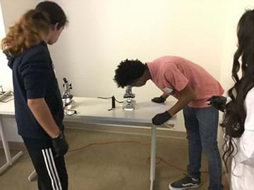
- Each team should collect their materials including: a microscope (with several light options: black light, base white light, or both), 2 pieces of graph paper, 4 slides, 4 slide cover slips, 4 onion samples, scalpel, tweezers, rubber gloves, and personal protection equipment.
- Have teams spend the first fifteen minutes looking over their materials, brainstorming and discussing which three imaging solutions they plan to test. Based on their ideas, students can draw out or write up their initial plan and develop the best strategies to image the cells on a piece of graph paper.
- After creating their plan, students first obtain their onion membrane samples. For best results, students should obtain a single layer of onion cells that allows light to easily pass through. To do so:
- Students carefully use the scalpel to slice one 2-cm onion strip apart and use tweezers find the membrane on the inside of the slice.
- Students then gently peel their cut onion sample until they isolate the membrane, resulting in one layer of onion that is super thin.
- Students place the onion membrane sample onto a clean slide, smoothing out any wrinkles with tweezers or the end of a pipette.
- These steps should be repeated for three more onion membrane samples
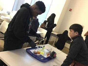
- Next, have students create a control by adding one or two drops of iodine solution onto the top of one onion membrane slide. Note: The iodine solution is used a as a control due to its common use in everyday cell slide imaging.
- After one minute, ask students to carefully drop the cover slip over the onion cells by placing one end of the cover slip into the iodine and dropping the other side down. This helps to prevent bubbles. (Note -- bubbles can be removed by lightly tapping with the bulb end of the pipette or by placing a tissue on the liquid at the edge of the cover slip close to the bubble.)
- Students repeat the procedure for each of their three chosen imaging solutions.
- After the four slides are ready, each team analyzes the control solution (iodine solution) slide by using the microscope to view the sample. Students should look for clarity of the structures and if they can see the cells and their organelles. Students write down what they observe and what the advantages and disadvantages are of the iodine sample on their graph paper.
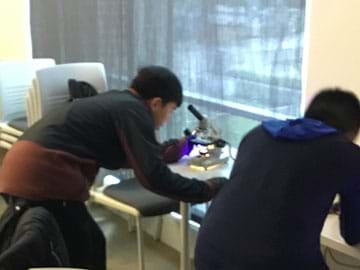
- Students next view the other samples under the microscope. Students should make notes and draw their observations of the sample on the graph paper. (Note -- if students used the fluorescent marker solution and/or the turmeric solution, any overhead lights need to be darkened.) Each team will then use the black light and shine it on the sample, pointing the light downward onto the slide.
- After viewing all their samples, have each team identifies which solution best images their onion membrane sample. Using that slide, students carefully, and in detail, draw the onion cells in the Onion Cell Lab Sheet. Students should label any organelles from in the sample. (Note -- the students should see the outline of the cell wall and possibly organelles within it.)
- While students work, have them think about the following investigating questions: What results do they expect? What worked? What didn’t work? Why didn’t it work? What would they do differently with the solutions? What else do they think would help make the onion cells more clear/easier to see?
Vocabulary/Definitions
cell: The basic structural and functional unit of living organisms.
cell membrane: A lipid barrier that encloses the cytoplasm and controls what enters and exits the cell.
cell wall: A tough, protective barrier that surrounds the outer membrane of some types of cells.
cytoplasm: The jelly-like material inside the outer membrane of a cell that holds the nucleus, organelles, and other components of the cell.
electromagnetic spectrum: A grouping of all possible energy levels of electromagnetic radiation from radio waves to gamma rays, including visible light.
fluorescent dye: A color dye that uses fluorescence of a certain substances to emit light. Used to identify cellular structures.
light waves: Electromagnetic waves with a frequency between ultraviolet and infrared waves, usually visible.
lissome : Membrane-bound organelles that contain enzymes capable of breaking down many types of molecules.
mitochondrion: An organelle in the cytoplasm of eukaryotic cells that functions in energy production.
nucleus: A membrane-bound structure in eukaryotic cells that contains the DNA.
organelle: A membrane-bound structure inside a cell that performs a specialized function.
ultraviolet waves: Electromagnetic waves with a shorter wavelength than visible light but longer than X-rays.
vacuole: A large, water filled organelle present in all plant and fungal cells and some animal and bacterial cells.
wavelength: The distance between any two corresponding points on successive oscillations of a wave, such as from peak to peak.
Assessment
Pre-Activity Assessment
Pre-Quiz: Have students complete the Pre-Activity Quiz.
Activity Embedded (Formative) Assessment
Lab Sheet: Have students complete the Onion Cell Lab Sheet.
Post-Activity (Summative) Assessment
Post-Quiz: Ask students the following questions:
- How do you know if a cell is a plant cell? (Answer: It has a cell wall.)
- What part of the electromagnetic spectrum does a fluorescent dye utilize? (Answer: Visible light waves.)
- Why do scientist use fluorescent dyes to view cell behavior? (Answer: To be able to see the cell’s biosynthesis at small measurements.)
- What does the nucleus do? (Answer: The nucleus controls the cells activities and carries the cell’s DNA.)
- What the role do cytoplast plays within the cell? (Answer: It helps with the cells metabolism and keys the organelles in place.)
- Out of all the methods to help image the cell, what method was the best? (Answer: This question is based on the students results of the activity. Though iodine solution is still the standard, some of the other dyes may be useful.)
Investigating Questions
These questions should be given before the lab.
- Can you see the onion cell and its organelles? (Answer: For most applications, the student should be able to see the cell wall and, possibly, the nucleus. The other organelles may be too small for the typical classroom microscope to pick up.)
- Does fluorescence help you clarify the samples and can you determine the cells organelles better? (Answer: The fluorescence should help with the cell imaging.)
- Why would a scientist use fluorescent dyes to identify cellular structures? (Answer: Should include the need for scientists to monitor the cells functions, fluorescent dyes help highlight images that are very small.)
Safety Issues
- Teacher should review with students how to properly handle scalpels and any other lab equipment used. Adult supervision should be constant throughout the lab.
- Fluorescent dye solution and iodine are not for human consumption and can be poisonous if ingested.
- Personal protection equipment, (lab coats or aprons, safety goggles, rubber gloves), need to be worn throughout lab to ensure safety of students and participants.
Troubleshooting Tips
Use pliers to pull apart the fluorescent marker-highlighters. Wear two gloves per hand as the marker dye may bleed through one set of rubber gloves.
Subscribe
Get the inside scoop on all things Teach Engineering such as new site features, curriculum updates, video releases, and more by signing up for our newsletter!More Curriculum Like This

Students learn how the heart functions. They are introduced to the concept of action potential generation, which causes the electrical current that triggers muscle contraction in the heart.
Copyright
© 2021 by Regents of the University of Colorado; original © 2020 Rice UniversityContributors
Robert Black, 8th Grade Science Teacher, Cleveland Middle School; Kim Calfee, 7th Grade Science Teacher, Cleveland Middle School; Ashley Boothe, Principal, Cleveland Middle SchoolSupporting Program
Nanotechnology Research Experience for Teachers , Rice UniversityAcknowledgements
This material was developed in collaboration with Rice University Office of STEM Engagement, based upon work supported by the National Science Foundation under grant no. NSF EEC-1711515—the Nanotechnology Research Experience for Teachers at the Rice University School Science and Technology in Houston, TX. Any opinions, findings and conclusions or recommendations expressed in this material are those of the author(s) and do not necessarily reflect the views of the National Science Foundation or Rice University.
Special thanks to
Dr. Han Xiao – Principal Investigator, Assistant Professor
Mentor: Dr. Juan Tang
Dr. Xiao’s Team
Rice University RET-STEM: Isaias Cerda, Christina Crawford & Carolyn Nichol
Last modified: September 2, 2021




User Comments & Tips