Quick Look
Grade Level: 10 (9-12)
Time Required: 45 minutes
Lesson Dependency:
Subject Areas: Biology
NGSS Performance Expectations:

| HS-LS1-1 |

Summary
Students are introduced to the latest imaging methods used to visualize molecular structures and the method of electrophoresis that is used to identify and compare genetic code (DNA). Students should already have basic knowledge of genetics, DNA (DNA structure, nucleotide bases), proteins and enzymes. The lesson begins with a discussion to motivate the need for imaging techniques and DNA analysis, which prepares students to participate in the associated two-part activity: 1) students each choose an imaging method to research (from a provided list of molecular imaging methods), 2) they research basic information about electrophoresis.Engineering Connection
Visualization of small structures such as the molecular structures of complex proteins and genetic material (DNA) is based on engineering discoveries and breakthroughs in physics at small scales. Imaging technologies such as x-ray and scanning electron microscopy—used by scientists and engineers to image microscopic structures—are also used by biomedical engineers and biologists to study biomolecules, cells and tissue samples. Microfluidics concepts and devices used to study colloidal particle flow are also employed by biologist to study and filter biomolecules. Gel electrophoresis is one example of the many engineering technologies that biologists use to compare fragments of DNA samples.
Learning Objectives
After this lesson, students should be able to:
- Enumerate some of the imaging technologies used for atomic scale microscopy.
- List the basic, underlying principles of the researched microscopy method.
- Describe how the microscopy method helped scientists to discover the structure of biomolecules.
- Explain the difference between molecular imaging and DNA gel electrophoresis.
- Explain that certain nucleotide base sequences in the DNA encode for proteins/enzymes, whereas the molecular shape of protein/enzyme determines their functions.
Educational Standards
Each Teach Engineering lesson or activity is correlated to one or more K-12 science,
technology, engineering or math (STEM) educational standards.
All 100,000+ K-12 STEM standards covered in Teach Engineering are collected, maintained and packaged by the Achievement Standards Network (ASN),
a project of D2L (www.achievementstandards.org).
In the ASN, standards are hierarchically structured: first by source; e.g., by state; within source by type; e.g., science or mathematics;
within type by subtype, then by grade, etc.
Each Teach Engineering lesson or activity is correlated to one or more K-12 science, technology, engineering or math (STEM) educational standards.
All 100,000+ K-12 STEM standards covered in Teach Engineering are collected, maintained and packaged by the Achievement Standards Network (ASN), a project of D2L (www.achievementstandards.org).
In the ASN, standards are hierarchically structured: first by source; e.g., by state; within source by type; e.g., science or mathematics; within type by subtype, then by grade, etc.
NGSS: Next Generation Science Standards - Science
| NGSS Performance Expectation | ||
|---|---|---|
|
HS-LS1-1. Construct an explanation based on evidence for how the structure of DNA determines the structure of proteins which carry out the essential functions of life through systems of specialized cells. (Grades 9 - 12) Do you agree with this alignment? |
||
| Click to view other curriculum aligned to this Performance Expectation | ||
| This lesson focuses on the following Three Dimensional Learning aspects of NGSS: | ||
| Science & Engineering Practices | Disciplinary Core Ideas | Crosscutting Concepts |
| Construct an explanation based on valid and reliable evidence obtained from a variety of sources (including students' own investigations, models, theories, simulations, peer review) and the assumption that theories and laws that describe the natural world operate today as they did in the past and will continue to do so in the future. Alignment agreement: | Systems of specialized cells within organisms help them perform the essential functions of life. Alignment agreement: All cells contain genetic information in the form of DNA molecules. Genes are regions in the DNA that contain the instructions that code for the formation of proteins, which carry out most of the work of cells.Alignment agreement: | Investigating or designing new systems or structures requires a detailed examination of the properties of different materials, the structures of different components, and connections of components to reveal its function and/or solve a problem. Alignment agreement: |
International Technology and Engineering Educators Association - Technology
-
The sciences of biochemistry and molecular biology have made it possible to manipulate the genetic information found in living creatures.
(Grades
9 -
12)
More Details
Do you agree with this alignment?
-
Connect technological progress to the advancement of other areas of knowledge and vice versa.
(Grades
9 -
12)
More Details
Do you agree with this alignment?
State Standards
Texas - Science
-
know that hypotheses are tentative and testable statements that must be capable of being supported or not supported by observational evidence. Hypotheses of durable explanatory power which have been tested over a wide variety of conditions are incorporated into theories;
(Grades
9 -
11)
More Details
Do you agree with this alignment?
-
know scientific theories are based on natural and physical phenomena and are capable of being tested by multiple independent researchers. Unlike hypotheses, scientific theories are well-established and highly-reliable explanations, but they may be subject to change as new areas of science and new technologies are developed;
(Grades
9 -
11)
More Details
Do you agree with this alignment?
-
distinguish between scientific hypotheses and scientific theories;
(Grades
9 -
11)
More Details
Do you agree with this alignment?
-
plan and implement descriptive, comparative, and experimental investigations, including asking questions, formulating testable hypotheses, and selecting equipment and technology;
(Grades
9 -
11)
More Details
Do you agree with this alignment?
-
analyze the levels of organization in biological systems and relate the levels to each other and to the whole system.
(Grades
9 -
11)
More Details
Do you agree with this alignment?
Pre-Req Knowledge
Basic knowledge about genetics: DNA, the four nucleotide bases and the base pairing rules, DNA double helix structure.
Introduction/Motivation
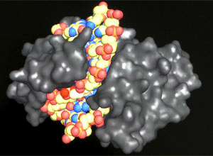
(Have ready to show students an assortment of molecular images of DNA, RNA, proteins and enzymes. See the Additional Multimedia Support section for a link to an online image database.)
Have you ever been curious about how your DNA might affect your chances of developing diseases and disorders? Do you ever wonder how scientists and doctors are able to study human DNA? Today we are going to learn about how engineering advances over decades have allowed scientists, doctors and other engineers to be able to visualize DNA and its structure.
The molecular structure of proteins and enzymes is very complex and plays a fundamental role in their functions. Slight changes in shape, known as protein folding, can result in anomalous function with adverse effects on the health of cells and of organisms. It has been recognized that many diseases and disorders are the result of protein/enzyme malfunctions; therefore, determining the structure of proteins and enzymes can help scientists and doctors develop more effective cures and treatments. Also, identifying which parts of the genetic code (DNA) are used as templates for the production of important proteins and enzymes is another major area of research interest. While the DNA structure is known, the important DNA segments that encode proteins are not entirely known and extensive research is performed to locate and identify them.
Genetics and the study of biomolecules, such as proteins and enzymes, rely in part on theoretical/computational models and on atomic scale microscopy. In particular, the discovery of the DNA structure—the double helix—and its replication and transcription processes has led to new discoveries in molecular biology and medicine. For years, scientists have tried to predict the arrangement of molecules (nucleotide bases, phosphate and sugar groups) that make up DNA using theoretical models based on the atomic and molecular interactions, but no validation or comparison between the structure predicted by models and the real structure existed. In 1953, the double helix structure of DNA based on x-ray analysis was published. A decade later, atomic force microscopy and other ultra-high resolution microscopy technologies were able to confirm this finding.
(Show students some molecular images of DNA, RNA, proteins and enzymes.) How have scientists been able to figure out the complex shapes of these tiny molecules? How do scientists know that the DNA or the hemoglobin look the way they do? Is it possible to look at the crystalline structure of molecules? The answer is yes, but not by using conventional microscopy. Instead, more complex technologies had to be invented, such as x-ray diffraction, transmission electron microscope (TEM), atomic force microscopy, fluorescence resonance energy transfer, magnetic resonance force microscopy, etc. What are these technologies? How do they work? What are the basic principles behind them? These questions all relate to modern molecular imaging. All these questions will be answered during this lesson and our discussion, and by your research (via the associated activity Inside the DNA).
Lesson Background and Concepts for Teachers

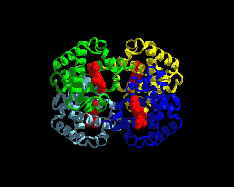
The molecular structure of chemical compounds and biological macromolecules (DNA, proteins) has been determined using x-rays, but now, newer imaging methods, such as electron microscopy and magnetic resonance, are being used as well. The structures of the DNA, RNA, proteins that you have seen in many images are schematics that show the approximated positions of the atoms that form particular molecules. For example, Figure 1 shows the molecular structure of hemoglobin, the blood protein responsible for oxygen transport. Each protein subunit is shown in a different color. The red subunits are hemes and are the locations where oxygen binds to hemoglobin. These schematics, as well as the crystalline structures of molecules, are made from data obtained by x-ray crystallography as well as theoretical models based on atomic and molecular interactions. It was only in the later part of the twentieth century that advancements in atomic scale microscopy enabled us to visualize molecular structures and validate many theoretical models.
The many types of molecular imaging technologies provide great information about molecular structures, but cannot provide information about the genetic code contained by the DNA or RNA. Molecular imaging cannot be used to compare two segments of DNA to tell if they are identical or not. Gel electrophoresis, a method based on the motility of polarized molecules in agarose gel when an electric current is a technology that can be used to compare DNA segments and determine their molecular weight.
This lesson and its associated activity make an excellent complement to the typical lesson on DNA structure or protein and enzyme structure/function. The lesson is designed to have students inquire about molecular imaging, the physics concepts behind it, and about DNA gel electrophoresis. As part of the lesson, include a discussion and presentation of the structure of biomolecules and the arrangement of atoms inside the molecules.
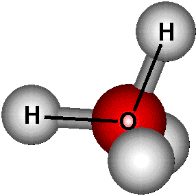
As a discussion starting point, present simple examples, such as the water molecule shown in Figure 2, with its three atoms arranged in a tetrahedron configuration. Proteins and enzymes are very complex molecules made of other molecules called subgroups, which are, in turn, made of amino acids, which, in turn, consist of smaller molecules. The interactions between all the molecules that make up a protein result in highly complex molecular structures.
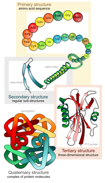
Based on the atomic and molecular interactions, four levels of molecular structure exist when describing the protein/enzyme structure (see Figure 3). The primary structure consists of the amino acid sequence that makes up a protein. The intermolecular interactions between the amino acids result in secondary structures that are further classified into beta sheets and alpha helices. The secondary structures also interact and result in more complex, tertiary and quaternary structures. The function of proteins/enzymes is highly dependent on the configuration of these structures and small alterations, such as adding, deleting or replacing one or more amino acids, can potentially result in defective proteins.
Conclude by introducing more complex molecular structures again, discussing the effect of atomic interactions of the molecular structures and how chemical models can predict the structure.


Associated Activities
- Inside the DNA - Students conduct research to learn about the engineering technology methods used by scientists to analyze or validate the molecular structure of DNA, proteins and enzymes, and basic information about electrophoresis and DNA identification. They share their findings through 10-slide class presentations.
Assessment
For a summary assessment of the lesson and its associated activity, see the Assessment section in the activity write-up for suggested criteria to use to evaluate student presentations on specific imaging technologies.
Additional Multimedia Support
Find protein and enzyme structure images at the RCSB Protein Data Bank database: http://www.rcsb.org/pdb/home/home.do
Subscribe
Get the inside scoop on all things Teach Engineering such as new site features, curriculum updates, video releases, and more by signing up for our newsletter!More Curriculum Like This

Students conduct their own research to discover and understand the methods designed by engineers and used by scientists to analyze or validate the molecular structure of DNA, proteins and enzymes, as well as basic information about gel electrophoresis and DNA identification.

Students learn how engineers apply their understanding of DNA to manipulate specific genes to produce desired traits, and how engineers have used this practice to address current problems facing humanity. Students fill out a flow chart to list the methods to modify genes to create GMOs and example a...

Students are introduced to genetic techniques such as DNA electrophoresis and imaging technologies used for molecular and DNA structure visualization. In the field of molecular biology and genetics, biomedical engineering plays an increasing role in the development of new medical treatments and disc...

Students focus on restriction enzymes and their applications to DNA analysis and DNA fingerprinting. They use this lesson and its associated activity in conjunction with biology lessons on DNA analysis and DNA replication.
Copyright
© 2013 by Regents of the University of Colorado; original © 2012 University of HoustonContributors
Mircea Ionescu; Myla Van DuynSupporting Program
University of Houston, National Science Foundation GK-12 and Research Experience for Teachers (RET) ProgramsAcknowledgements
This digital library content was developed by the University of Houston's College of Engineering under National Science Foundation GK-12 grant number DGE-0840889. However, these contents do not necessarily represent the policies of the NSF and you should not assume endorsement by the federal government.
Last modified: June 14, 2019


User Comments & Tips