Quick Look
Grade Level: 10 (9-12)
Time Required: 45 minutes
Expendable Cost/Group: US $0.00
Group Size: 1
Activity Dependency:
Subject Areas: Biology
NGSS Performance Expectations:

| HS-LS1-1 |

Summary
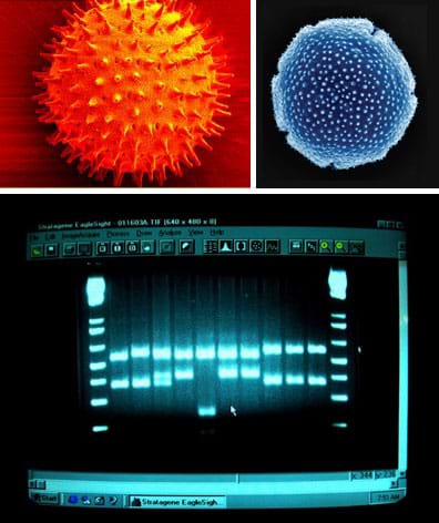
Students conduct their own research to discover and understand the methods designed by engineers and used by scientists to analyze or validate the molecular structure of DNA, proteins and enzymes, as well as basic information about gel electrophoresis and DNA identification. In this computer-based activity, students investigate particular molecular imaging technologies, such as x-ray, atomic force microscopy, transmission electron microscopy, and create short PowerPoint presentations that address key points. The presentations include their own explanations of the difference between molecular imaging and gel electrophoresis.Engineering Connection
Visualization of small structures such as molecular structures of complex proteins and genetic material (DNA) is based on engineering discoveries and breakthroughs in physics at small scales. Imaging technologies such as x-ray and scanning electron microscopy—used in by scientists and engineers to image microscopic structures—are also used by biomedical engineers and biologists to study biomolecules, cells and tissue samples.
Learning Objectives
After this activity, students should be able to:
- Enumerate some of the imaging technologies used for atomic scale microscopy.
- List the basic, underlying principles of the researched microscopy method.
- Describe how the microscopy method helped scientists to discover the structure of biomolecules.
- Explain the difference between molecular imaging and DNA gel electrophoresis.
- Explain that certain nucleotide base sequences in the DNA encode for proteins/enzymes whereas the molecular shape of protein/enzyme determines their function.
Educational Standards
Each Teach Engineering lesson or activity is correlated to one or more K-12 science,
technology, engineering or math (STEM) educational standards.
All 100,000+ K-12 STEM standards covered in Teach Engineering are collected, maintained and packaged by the Achievement Standards Network (ASN),
a project of D2L (www.achievementstandards.org).
In the ASN, standards are hierarchically structured: first by source; e.g., by state; within source by type; e.g., science or mathematics;
within type by subtype, then by grade, etc.
Each Teach Engineering lesson or activity is correlated to one or more K-12 science, technology, engineering or math (STEM) educational standards.
All 100,000+ K-12 STEM standards covered in Teach Engineering are collected, maintained and packaged by the Achievement Standards Network (ASN), a project of D2L (www.achievementstandards.org).
In the ASN, standards are hierarchically structured: first by source; e.g., by state; within source by type; e.g., science or mathematics; within type by subtype, then by grade, etc.
NGSS: Next Generation Science Standards - Science
| NGSS Performance Expectation | ||
|---|---|---|
|
HS-LS1-1. Construct an explanation based on evidence for how the structure of DNA determines the structure of proteins which carry out the essential functions of life through systems of specialized cells. (Grades 9 - 12) Do you agree with this alignment? |
||
| Click to view other curriculum aligned to this Performance Expectation | ||
| This activity focuses on the following Three Dimensional Learning aspects of NGSS: | ||
| Science & Engineering Practices | Disciplinary Core Ideas | Crosscutting Concepts |
| Construct an explanation based on valid and reliable evidence obtained from a variety of sources (including students' own investigations, models, theories, simulations, peer review) and the assumption that theories and laws that describe the natural world operate today as they did in the past and will continue to do so in the future. Alignment agreement: | Systems of specialized cells within organisms help them perform the essential functions of life. Alignment agreement: All cells contain genetic information in the form of DNA molecules. Genes are regions in the DNA that contain the instructions that code for the formation of proteins, which carry out most of the work of cells.Alignment agreement: | Investigating or designing new systems or structures requires a detailed examination of the properties of different materials, the structures of different components, and connections of components to reveal its function and/or solve a problem. Alignment agreement: |
International Technology and Engineering Educators Association - Technology
-
Students will develop an understanding of the relationships among technologies and the connections between technology and other fields of study.
(Grades
K -
12)
More Details
Do you agree with this alignment?
-
The sciences of biochemistry and molecular biology have made it possible to manipulate the genetic information found in living creatures.
(Grades
9 -
12)
More Details
Do you agree with this alignment?
-
Connect technological progress to the advancement of other areas of knowledge and vice versa.
(Grades
9 -
12)
More Details
Do you agree with this alignment?
State Standards
Texas - Science
-
analyze the levels of organization in biological systems and relate the levels to each other and to the whole system.
(Grades
9 -
11)
More Details
Do you agree with this alignment?
-
identify components of DNA, and describe how information for specifying the traits of an organism is carried in the DNA;
(Grades
9 -
11)
More Details
Do you agree with this alignment?
-
describe how techniques such as DNA fingerprinting, genetic modifications, and chromosomal analysis are used to study the genomes of organisms.
(Grades
9 -
11)
More Details
Do you agree with this alignment?
Materials List
Each student needs:
- computer with Internet connection, for research work
- PowerPoint software, for preparing slide presentations
Pre-Req Knowledge
Basic knowledge about genetics: DNA, the four nucleotide bases and the base pairing rules, DNA double helix structure.
Introduction/Motivation
(This activity follows the associated lesson, so first present the lesson's Introduction/Motivation section, followed by a class discussion on the related bioscience, as provided in the lesson's Teacher Background section. Then proceed with the following Introduction/Motivation and the research activity.)
What are the methods currently used by scientists to analyze or validate the molecular structure of DNA, proteins and enzymes? How have scientists figured out the complex shapes of DNA or hemoglobin? How do we know what they look like? Is it possible to look at the crystalline structure of molecules? (Listen to student ideas gleaned from the associated lesson.) Using conventional microscopes is not enough to see at the atomic scale. Instead, engineers and scientists have devised more complex technologies. Some examples of molecular imaging methods include: x-ray diffraction, transmission electron microscope (TEM), atomic force microscopy, fluorescence resonance energy transfer, magnetic resonance force microscopy, etc. These molecular imaging technologies are able to provide information about the structures.
If we want to know more, molecular imaging is unable to provide information about the genetic code contained by the DNA or RNA. These various imaging methods do not provide information about the content of the DNA, that is, the particular sequences of nucleotide bases (adenine, thymine, cytosine and guanine). And, molecular imaging cannot be used to compare two segments of DNA to tell if they are identical or not. In order to analyze the content of DNA segments or to compare them requires a different approach than just visualizing the DNA structure.
For that task, gel electrophoresis is a relatively simple and inexpensive method designed to analyze DNA molecules and determine their sequences. Gel electrophoresis is based on the motility of polarized molecules in agarose gel when an electric current is applied. To do this, DNA segments are introduced in a gel solution and the segments travel through the gel as en electric current is applied. Depending on size and composition, the DNA segments travel at different speeds (very low speeds) with similar segments traveling at similar rates. So gel electrophoresis is way to analyze and compare DNA segments without looking at the DNA's actual molecular structure. With gel electrophoresis, scientists can compare DNA segments and determine their molecular weight.
So what is the difference between these two methods: molecular imaging and gel electrophoresis? (Listen to student responses to gauge their understanding.) Both methods are used to analyze proteins, enzymes and genetic molecules (DNA, RNA), and are usually used in conjunction. Molecular imaging is used to determine molecular structure, while gel electrophoresis is used to determine molecular composition.
We've mentioned many different molecular imaging technologies. But how do they work? What are the basic principles behind them? Let's find out more.
Procedure
With the Students
- Present to the class the content of the Introduction/Motivation section.
- Research Part 1: From the list below, assign (or let students choose) microscopy technologies to research. The list includes suggested online resources to get them started. Direct students to build presentations from their findings. Inform them of the required presentation components (see the Assessment section.) Give students time to conduct the internet research and compose a slide presentation.
- x-ray crystallography https://en.wikipedia.org/wiki/X-ray_crystallography
- transmission electron microscope (TEM) https://en.wikipedia.org/wiki/Transmission_electron_microscopy_DNA_sequencing
- atomic force microscopy (AFM) http://www.pnas.org/content/94/2/496.short
- fluorescence resonance energy transfer (FRET) https://en.wikipedia.org/wiki/F%C3%B6rster_resonance_energy_transfer
- magnetic resonance force microscopy https://www.nytimes.com/2009/01/13/science/13mri.html?_r=2&
- photo activated localization microscopy (PALM) imaging https://en.wikipedia.org/wiki/Photoactivated_localization_microscopy
- Research Part 2: Have students look for information on DNA gel electrophoresis and each prepare a slide describing how it works and its application to DNA analysis and comparison. A useful place to start researching about DNA electrophoresis is: https://www.jove.com/science-education/5057/dna-gel-electrophoresis
- Presentations: Have students give their presentations to the rest of the class. If time is limited, at a minimum allow time for one presentation on each of the seven different technologies, so the rest of the class learns about all of them. Have all students turn in their presentations to the teacher for grading. Evaluate each student's presentation based on meeting the criteria listed in the Assessment section.
Vocabulary/Definitions
crystalline structure: A unique arrangement of atoms or molecules in a crystalline liquid or solid.
DNA: Acronym for deoxyribonucleic acid. A self-replicating material present in nearly all living organisms as the main constituent of chromosomes.
protein: Any of a group of complex organic macromolecules that contain carbon, hydrogen, oxygen, nitrogen and usually sulfur, and are composed of one or more chains of amino acids.
RNA: Acronym for ribonucleic acid. A nucleic acid present in all living cells. Its principal role is to act as a messenger carrying instructions from DNA for controlling the synthesis of proteins.
Assessment
Post-Activity Assessment
Research Presentations: Evaluate student presentations of their imaging research findings as a summative assessment for the activity and its associated lesson. Challenge students to be clear and concise, providing the required information in no more than 10 slides. Require each presentation to contain the following components, and evaluate accordingly.
- date when the method/technology was invented
- physical phenomena involved (how it works, for example: electron scattering, nanosized probe/detector, resonant frequency, without providing too many details; summarize the basic concepts)
- spatial resolution (the size of the smallest object that can be observed)
- engineering and technical challenges and break-throughs (such as the design of special detectors or microscopic probes)
- accomplishments in imaging DNA/proteins (what it has enabled scientists to discover/see)
- example images of DNA, proteins or other biological macromolecules obtained with the visualization method
- description of DNA gel electrophoresis technology
- explanation of the difference between molecular imaging and DNA electrophoresis
- list of sources and their URLs
Subscribe
Get the inside scoop on all things Teach Engineering such as new site features, curriculum updates, video releases, and more by signing up for our newsletter!More Curriculum Like This

Students are introduced to the latest imaging methods used to visualize molecular structures and the method of electrophoresis that is used to identify and compare genetic code (DNA).

Students are introduced to genetic techniques such as DNA electrophoresis and imaging technologies used for molecular and DNA structure visualization. In the field of molecular biology and genetics, biomedical engineering plays an increasing role in the development of new medical treatments and disc...

Students focus on restriction enzymes and their applications to DNA analysis and DNA fingerprinting. They use this lesson and its associated activity in conjunction with biology lessons on DNA analysis and DNA replication.

Students learn how engineers apply their understanding of DNA to manipulate specific genes to produce desired traits, and how engineers have used this practice to address current problems facing humanity. Students fill out a flow chart to list the methods to modify genes to create GMOs and example a...
Copyright
© 2013 by Regents of the University of Colorado; original © 2012 University of HoustonContributors
Mircea Ionescu; Myla Van DuynSupporting Program
National Science Foundation GK-12 and Research Experience for Teachers (RET) Programs, University of HoustonAcknowledgements
This digital library content was developed by the University of Houston's College of Engineering under National Science Foundation GK-12 grant number DGE 0840889. However, these contents do not necessarily represent the policies of the NSF and you should not assume endorsement by the federal government.
Last modified: March 29, 2022


User Comments & Tips