Quick Look
Summary
Students learn about the parts of the human respiratory system and the gas exchange process that occurs in the lungs. They also learn about the changes in the respiratory system that occur during spaceflight, such as decreased lung capacity.Engineering Connection
By studying the respiratory system, engineers have been able to come up with assistive technology, such as the heart-lung machine. This machine is hooked up to the blood vessels that lead to and from the heart, allowing blood to be pumped into the machine to receive oxygen and get rid of carbon dioxide (such as what the lungs would normally do). The machine accomplishes this by directing the blood through a series of chambers before diverting it back into the body. This invention is a critical part of life-saving surgeries that involve organ transplants (usually the heart). Chemical engineers develop pharmaceuticals that can aid lung diseases, such as emphysema, pneumonia, cystic fibrosis, and asthma. Engineers are currently working on creating an implantable, artificial lung that would aid patients with serious lung disease.
Learning Objectives
After this lesson, students should be able to:
- Identify and describe the components of the respiratory system.
- Describe what happens during the inhalation and exhalation process.
- Describe how the respiratory system is affected during space flight.
- List several innovations that engineers have designed related to the respiratory system.
Educational Standards
Each Teach Engineering lesson or activity is correlated to one or more K-12 science,
technology, engineering or math (STEM) educational standards.
All 100,000+ K-12 STEM standards covered in Teach Engineering are collected, maintained and packaged by the Achievement Standards Network (ASN),
a project of D2L (www.achievementstandards.org).
In the ASN, standards are hierarchically structured: first by source; e.g., by state; within source by type; e.g., science or mathematics;
within type by subtype, then by grade, etc.
Each Teach Engineering lesson or activity is correlated to one or more K-12 science, technology, engineering or math (STEM) educational standards.
All 100,000+ K-12 STEM standards covered in Teach Engineering are collected, maintained and packaged by the Achievement Standards Network (ASN), a project of D2L (www.achievementstandards.org).
In the ASN, standards are hierarchically structured: first by source; e.g., by state; within source by type; e.g., science or mathematics; within type by subtype, then by grade, etc.
NGSS: Next Generation Science Standards - Science
-
CCC.4.3-5.2.
A system can be described in terms of its components and their interactions.
(Grades 3 - 5)
More Details
Do you agree with this alignment?
-
CCC.8.3-5.8.
Engineers improve existing technologies or develop new ones to increase their benefits, to decrease known risks, and to meet societal demands.
(Grades 3 - 5)
More Details
Do you agree with this alignment?
International Technology and Engineering Educators Association - Technology
-
Technological advances have made it possible to create new devices, to repair or replace certain parts of the body, and to provide a means for mobility.
(Grades
3 -
5)
More Details
Do you agree with this alignment?
-
Describe how a subsystem is a system that operates as part of another, larger system.
(Grades
3 -
5)
More Details
Do you agree with this alignment?
State Standards
Colorado - Science
-
Human body systems have basic structures, functions, and needs
(Grade
5)
More Details
Do you agree with this alignment?
Pre-Req Knowledge
A basic understanding of red blood cells (Lesson 5) is necessary for this lesson.
Introduction/Motivation
Did you know that you can live for about 30 days without food, three days without water, but only three minutes without air! You breathe in and out 15-25 times (approximately 250 ml of oxygen and 200 ml of carbon dioxide) per minute without even thinking about it! Every organ in our bodies needs oxygen in order to function properly and keep us alive. The respiratory system delivers this precious oxygen to our blood that then delivers it throughout our bodies, just as we learned in the circulatory system. The harder your body is working (exercising), the more oxygen it needs, which causes you to breathe harder and faster.
The respiratory system is made of trachea, bronchi, alveoli and the lungs. These organs are protected by your rib cage, which helps us breathe by moving as we breathe in and out. When we breathe in, the rib cage swings up and out: making more space for air to enter the lungs. When we breathe out, the rib cage moves down and back: pushing air from the lungs out the nose and mouth.
Sometimes people have lungs that do not work properly. Many lung diseases can affect how well a person is able to breathe, such as asthma, emphysema, bronchitis and pneumonia. By studying the respiratory system and lung diseases, chemical engineers can develop medicines to treat these diseases (such as inhalers for asthmatics). Engineers are also working on developing an artificial lung that would help people to live long enough to fight off potentially deadly infections. Currently, however, the artificial lung is only suitable to use for about two weeks. The artificial lung is approximately 18 inches long and consists of membranes that pass oxygen to the blood and remove carbon dioxide. The way in which the artificial lung is inserted is very interesting: it is inserted through a vein in the leg and lodged in the main vein (the vena cavae), passing blood to the heart. The blood is re-oxygenated through a catheter that is attached to an oxygen supply.
What happens to an astronaut's lung when traveling in space? Can you guess? Can a person breathe in space with out any help? No, they cannot, because there is no air in space. The respiratory system needs oxygen and pressure to push the air in and out of the lungs. This is where engineers can help.
Inside a spacecraft, engineers have designed systems to provide the right amount of oxygen and air pressure — similar to the air and pressure provided while flying in an airplane. The body's respiratory system is probably the least affected during spaceflight as long, provided there is plenty of oxygen available and the air pressure inside the spacecraft remains at an appropriate level. This is the same for the specially engineered space suits that astronauts have worn when they walk on the moon. The main difference when breathing in space is that there is a decrease in lung capacity — meaning astronauts cannot take in as much oxygen in a single breath as they can on Earth. Due to weightlessness, the organs in the abdominal cavity are no longer pulled downward causing them to shift up and press on the diaphragm. This decreases how much air the lungs can take in during a single breath. Although astronauts cannot inhale as much air at one time, this change is quickly reversed once they return to Earth.
Today, we are going to learn more about how the respiratory system works and how engineers help people with respiratory problems and help astronauts travel in space. Following the lesson students will have the opportunity to further investigate the respiration process by creating their own model lung with the associated activity Creating Model Working Lungs: Just Breathe.
Lesson Background and Concepts for Teachers
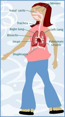
The respiratory system consists of the trachea, bronchi, alveoli, and the lungs and is responsible for gas exchange between the environment and the body (delivery of oxygen from the lungs to the bloodstream and elimination of carbon dioxide from the bloodstream to the lungs). This system — along with the heart (located between the lungs; in fact, the left lung is slightly smaller to accommodate the heart) — is located in the thoracic cavity of the ribcage that provides protection. The diaphragm muscle separates this system from the abdominal cavity. See Figure 2 for an illustration of the chest cavity.
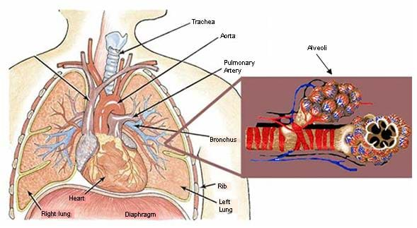
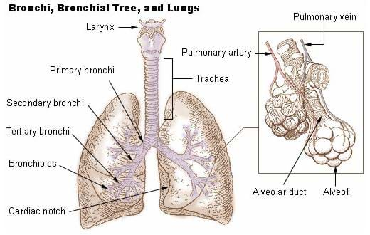
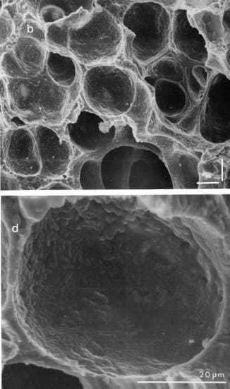
Air first enters the body through the nose or mouth and then travels down the throat to the trachea (windpipe). Rings of cartilage protect the trachea from collapsing and subsequently blocking air from entering the lungs. The trachea then splits into two large tubes (one for each lung) called the right and left bronchi. The bronchi split into smaller bronchi and eventually become tiny bronchioles inside the lungs (see Figure 3). These bronchioles end in clusters of alveoli, which are tiny, thin-walled, air sacs that inflate as air enters the lungs and deflate as air exits (see Figure 4). The average, adult lung contains approximately 300 millions of these expandable sacs. Gas exchange then occurs between the alveoli and the pulmonary capillaries (tiny blood vessels) that lie within the alveoli walls. During inhalation, oxygen travels from the alveoli (high oxygen concentration) to the blood (lower oxygen concentration). Oxygen is then stored in the hemoglobin of the red blood cells to be delivered to the rest of the body. Conversely, during exhalation, carbon dioxide travels from the blood to the air.
During inhalation, the diaphragm contracts downward and rib muscles pull upward causing air to fill the lungs (increases the volume of the thoracic cavity and decreases pressure in the lungs so the air will flow from the higher-pressure environment to lower-pressure area in the lungs). The diaphragm then relaxes, and the lungs contract which causes air to be pushed out from the lungs (exhalation).
Some common types of lung disease are:
- Asthma: When respiratory muscles have to work harder due to the bronchioles constricting, which creates smaller airways (see Figure 5). It is a response to an allergen or other irritant (such as animal fur).
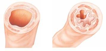
- Emphysema: When respiratory muscles have to work harder due to the fact that the lungs have become stiff with fibers and are less elastic in nature.
- Bronchitis: When respiratory muscles have to work harder due to inflamed, narrow airways.
- Pulmonary Edema: A slowing of the gas exchange due to a build up of fluid between the alveolus and pulmonary capillary (increases distance that the gases have to travel).
- Pneumonia: Bacteria or virus that affects the membrane (pleura) that surrounds the lungs.
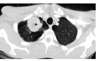
- Lung Cancer: When malignant cells divide in the lung tissue, possibly invading nearby tissues or spreading through the blood stream; oftentimes caused by smoking. Figure 6 illustrates the presence of cancer in the lungs: the black areas are the lungs and the white patch on the right lung is cancer.
Associated Activities
- Creating Model Working Lungs: Just Breathe - Students build model lungs to investigate the process of inhalation and exhalation. They discuss what happens during respiration and what might happen if the respiratory system were damaged.
Lesson Closure
The respiratory system is closely linked with the circulatory system (they are even sometimes combined into the cardiovascular system since the heart, blood and lungs work together to provide oxygen to the body). The ability for air to reach the lungs and the lungs to deliver oxygen to the bloodstream (and elimination of carbon dioxide from the bloodstream to the lungs) is vital to the survival of the rest of the systems of our body. Without breathing, our whole body would shut down from lack of oxygen in the bloodstream. It is a good thing we breathe without having to remember or even to think about it!
Who remembers the organs in the respiratory system? (Answer: The trachea, bronchi, alveoli and lungs.) These are protected by your rib cage, which also helps us breathe by moving in and out. Place your hand over your ribcage. Can you feel it moving in and out as your breath?
How do engineers help people who have respiratory problems? Chemical engineers have created medicines for people with asthma, emphysema, bronchitis and pneumonia. Engineers have also designed an artificial lung to help people fight off infections. Lastly, engineers help design systems to give astronauts the right amount of air pressure and oxygen to survive in space!
Vocabulary/Definitions
alveoli: Terminal sacs where gas exchange occurs; they constitute the majority of the lung tissue and give the lungs a soft, spongy texture.
aveolar duct: Tiny tubules at the ends of bronchioles that divide into alveolar sacs.
bronchi: The two large tubes connected to the trachea that carry air to and from the lungs.
bronchiole: Branches of the bronchi which distribute air to the alveoli (smaller than one millimeter in diameter); they are the first airway passages that no longer contain cartilage.
cartilage: A type of dense connective tissue.
diaphragm: A shelf of muscle extending across the bottom of the ribcage.
intercostal muscle: Muscles that line the rib cage and assist in breathing.
lungs: Spongy, saclike respiratory organs that occupy the chest cavity (with the heart) and provide oxygen to the blood and remove carbon dioxide from it.
pleural membrane: Double-layered membranes that cover the lungs, separating them from other organs (forms a fluid-filled chest cavity in order to reduce friction during inhalation and exhalation).
pulmonary capillary: A tiny blood vessel that surround each alveolus.
trachea: The air tube that connects with the bronchi; lined with ciliated (finger-like projections) cells to keep foreign bodies out and reinforced with cartilage rings to keep it from collapsing. Also called the windpipe.
Assessment
Pre-Lesson Assessment
Discussion Question: Solicit, integrate and summarize student responses.
- Ask the students if they know what happens inside their body when they breathe in and out. Do you breathe faster or slower when you are playing a sport? Why? (Answer: You breathe faster, because your body is using more oxygen.)
Post-Introduction Assessment
Voting: Ask a true/false question and have students vote by holding thumbs up for true and thumbs down for false. Count the votes and write the totals on the board. Give the right answer.
- True or False: The lungs are part of the respiratory system. (Answer: True)
- True or False: Our rib cage pushes in when we breathe in. (Answer: False. When we breathe in, our rib cage swings up and out, making more space for air to enter our lungs; when we breathe out, our rib cage moves down and back, pushing air from our lungs out our nose and mouth.)
- True or False: An astronaut can breathe easily in space the same as they do on Earth. (Answer: False. An astronaut cannot breathe the same in space, because there is no air in space. The respiratory system needs oxygen and pressure to push the air in and out of the lungs.)
- True or False: Engineers design devices to help astronauts breathe in space. (Answer: True. Engineers have designed systems to provide the right air pressure and oxygen amount during space flight and space exploration.)
- True or False: Engineers work to help people who have respiratory problems (difficulty breathing properly). (Answer: True. Chemical engineers have created medicines for people with asthma, emphysema, bronchitis and pneumonia. Engineers have also designed an artificial lung to help people fight off infections. Lastly, engineers help design systems to give astronauts the right amount of air pressure and oxygen to survive in space.)
Lesson Summary Assessment
Send-a-Problem: Have students write their own questions about the respiratory system. Each student on a team creates a flashcard with a question on one side and the answer on the other. If the team cannot agree on an answer they should consult the teacher. Pass the flashcards to the next team. Each member of the team reads a flashcard and everyone attempts to answer it. If they are right, they pass the card on to another team. If they feel they have another correct answer, they can write it answer on the back of the flashcard as an alternative answer. Once all teams have tested themselves on all the flashcards, clarify any questions.
Thoughtful Questions: Ask the students the following extension questions about the respiratory system
- Does the way you breathe change during space flight? (Answer: No, but your lung capacity decreases due to the fact that other internal organs shift upward in microgravity.)
- Can people breathe underwater? (Answer: People can only breathe underwater if they take an air supply with them. Engineers have helped design air supply devices, such as diving tanks, to help people breathe in extreme conditions.)
Lesson Extension Activities
Have students investigate the diaphragm and how it used when we breathe. How do they think the diaphragm changes during space flight? (Answer: The muscles shrink in a microgravity environment.) Relate this back to any lessons on the muscular system (such as Lesson 2 of the unit). How might engineers help astronauts have a healthy diaphragm in space? The following National Space Biomedical Research Institute website has a number of student investigations that describe some of the physiological changes that astronauts go through during space flight. They are mostly geared towards older students but may provide some ideas for younger learners: http://www.nsbri.org/HumanPhysSpace/
Subscribe
Get the inside scoop on all things Teach Engineering such as new site features, curriculum updates, video releases, and more by signing up for our newsletter!More Curriculum Like This

Students are introduced to the respiratory system, the lungs and air. They learn about how the lungs and diaphragm work, how air pollution affects lungs and respiratory functions, some widespread respiratory problems, and how engineers help us stay healthy by designing machines and medicines that su...

To gain a better understanding of the roles and functions of components of the human respiratory system and our need for clean air, students construct model lungs that include a diaphragm and chest cavity. Student teams design and build a prototype face mask pollution filter and use their model lung...

This lesson describes how the circulatory system works, including the heart, blood vessels and blood. Students learn about the chambers and valves of the heart, the difference between veins and arteries, and the different components of blood.

Students explore the inhalation/exhalation process that occurs in the lungs during respiration. Using everyday materials, each student team creates a model pair of lungs.
References
BBC News, Heath, "Artificial lung breakthrough," April 26, 2001. www.bbc.co.uk/news Accessed May 23, 2006
Lujan, Barbara. and White, Ronald. National Space Biomedical Research Institute, Human Physiology in Space, Houston, TX: GPO, 1995. www.nsbri.org Accessed May 23, 2006
Short, Sr., Nicholas M. National Aeronautics and Space Administration, Information Sciences Branch, Goddard Space Flight Center, The Remote Sensing Tutorial (RST).
U.S. Department of Health and Human Services, Office on Women's Health, National Women's Health Information Center (NWHIC), GirlsHealth.gov, Body – Becoming a Woman, "Learn about your whole body – from your heart to your bones," March 2006.
U.S. Department of Health and Human Services, Office on Women's Health, National Women's Health Information Center (NWHIC), Lung Disease, March 2006.
U.S. National Cancer Institute's Surveillance, Epidemiology and End Results (SEER) Program, Bronchi, Bronchial Tree, and Lungs, "Bronchi and Bronchial Tree."
Copyright
© 2006 by Regents of the University of ColoradoContributors
Teresa Ellis; Denali Lander; Malinda Schaefer Zarske; Janet YowelSupporting Program
Integrated Teaching and Learning Program, College of Engineering, University of Colorado BoulderAcknowledgements
The contents of this digital library curriculum were developed under grants from the Fund for the Improvement of Postsecondary Education (FIPSE), U.S. Department of Education and National Science Foundation (GK-12 grant no. 0338326). However, these contents do not necessarily represent the policies of the Department of Education or National Science Foundation, and you should not assume endorsement by the federal government.
Last modified: March 12, 2022


User Comments & Tips