Quick Look
Grade Level: 10 (11-12)
Time Required: 6 hours 45 minutes
(eight 50-minute sessions)
Expendable Cost/Group: US $0.00
Group Size: 3
Activity Dependency: None
Subject Areas: Biology, Chemistry, Computer Science, Data Analysis and Probability, Life Science, Measurement, Problem Solving, Science and Technology
NGSS Performance Expectations:

| HS-ETS1-3 |
| HS-ETS1-4 |
Summary
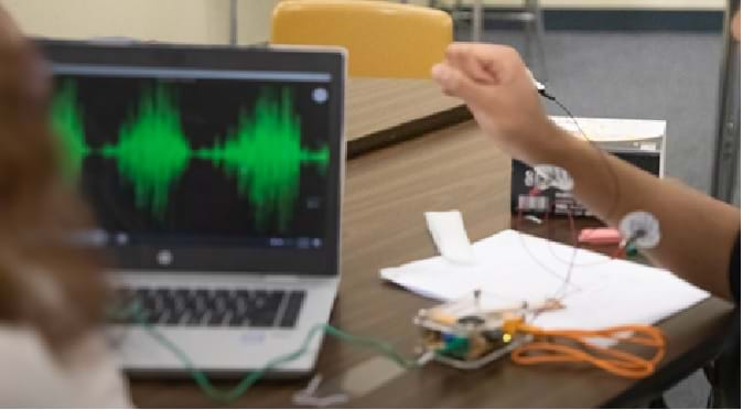
In this second activity, students dive deeper into the neuromuscular system by exploring how the body recruits and activates muscles in response to various gestures. They begin by examining the neuromuscular junction and diagramming the neuronal circuitry pathway involved in muscle activation. Using electromyography (EMG), students learn how to measure subtle muscle responses triggered by wrist and finger movements. They collect and analyze EMG data with the help of surface electrodes and computer software, focusing on recording and processing signals from both large and small muscle groups at different speeds. By practicing data acquisition, filtering, and signal analysis, students apply the scientific method and engineering design process to understand how neurons interact with muscles. As they work in teams to compare signal parameters such as amplitude and frequency, they gain valuable insights into muscle recruitment and learn to distinguish between different types of gestures based on their EMG data. This activity reinforces students' understanding of neuromuscular function while enhancing their skills in data collection and analysis.Engineering Connection
EMG technology draws on multiple engineering disciplines. Biomedical engineers develop and refine EMG sensors and signal processing methods. Electrical engineers design circuits and systems to acquire, filter, and interpret muscle signals. Mechanical engineers study muscle biomechanics to guide sensor placement and understand force dynamics. Computer engineers create software and algorithms—often using machine learning—for data analysis and gesture recognition. Neuroengineers, a branch of biomedical engineering, focus on neuron-muscle interactions to optimize EMG for medical, prosthetic, and rehab applications. Together, these disciplines drive EMG innovation across healthcare, robotics, and human-computer interaction.
Learning Objectives
After this activity, students should be able to:
- Describe muscle physiology and neuron/muscle communication using electromyograms (EMG), specifically explain EMG signal property changes between large and fine muscle.
- Acquire and analyze biological data to make valid and reliable scientific observations.
- Discuss, hypothesize, and suggest other experiments based on learning from these EMG studies (e.g., carpal tunnel syndrome, Parkinson’s to robotic/neuroprosthetic applications).
Educational Standards
Each TeachEngineering lesson or activity is correlated to one or more K-12 science,
technology, engineering or math (STEM) educational standards.
All 100,000+ K-12 STEM standards covered in TeachEngineering are collected, maintained and packaged by the Achievement Standards Network (ASN),
a project of D2L (www.achievementstandards.org).
In the ASN, standards are hierarchically structured: first by source; e.g., by state; within source by type; e.g., science or mathematics;
within type by subtype, then by grade, etc.
Each TeachEngineering lesson or activity is correlated to one or more K-12 science, technology, engineering or math (STEM) educational standards.
All 100,000+ K-12 STEM standards covered in TeachEngineering are collected, maintained and packaged by the Achievement Standards Network (ASN), a project of D2L (www.achievementstandards.org).
In the ASN, standards are hierarchically structured: first by source; e.g., by state; within source by type; e.g., science or mathematics; within type by subtype, then by grade, etc.
NGSS: Next Generation Science Standards - Science
| NGSS Performance Expectation | ||
|---|---|---|
|
HS-ETS1-3. Evaluate a solution to a complex real-world problem based on prioritized criteria and trade-offs that account for a range of constraints, including cost, safety, reliability, and aesthetics, as well as possible social, cultural, and environmental impacts. (Grades 9 - 12) Do you agree with this alignment? |
||
| Click to view other curriculum aligned to this Performance Expectation | ||
| This activity focuses on the following Three Dimensional Learning aspects of NGSS: | ||
| Science & Engineering Practices | Disciplinary Core Ideas | Crosscutting Concepts |
| Evaluate a solution to a complex real-world problem, based on scientific knowledge, student-generated sources of evidence, prioritized criteria, and tradeoff considerations. Alignment agreement: | When evaluating solutions it is important to take into account a range of constraints including cost, safety, reliability and aesthetics and to consider social, cultural and environmental impacts. Alignment agreement: | New technologies can have deep impacts on society and the environment, including some that were not anticipated. Analysis of costs and benefits is a critical aspect of decisions about technology. Alignment agreement: |
| NGSS Performance Expectation | ||
|---|---|---|
|
HS-ETS1-4. Use a computer simulation to model the impact of proposed solutions to a complex real-world problem with numerous criteria and constraints on interactions within and between systems relevant to the problem. (Grades 9 - 12) Do you agree with this alignment? |
||
| Click to view other curriculum aligned to this Performance Expectation | ||
| This activity focuses on the following Three Dimensional Learning aspects of NGSS: | ||
| Science & Engineering Practices | Disciplinary Core Ideas | Crosscutting Concepts |
| Use mathematical models and/or computer simulations to predict the effects of a design solution on systems and/or the interactions between systems. Alignment agreement: | Both physical models and computers can be used in various ways to aid in the engineering design process. Computers are useful for a variety of purposes, such as running simulations to test different ways of solving a problem or to see which one is most efficient or economical; and in making a persuasive presentation to a client about how a given design will meet his or her needs. Alignment agreement: | Models (e.g., physical, mathematical, computer models) can be used to simulate systems and interactions—including energy, matter, and information flows—within and between systems at different scales. Alignment agreement: |
-
SEP.2.9-12.3.
Analyze complex real-world problems by specifying criteria and constraints for successful solutions.
(Grades 9 - 12)
More Details
Do you agree with this alignment?
Materials List
For the entire class to share:
- laptop or computer with Internet access and a projector (to show YouTube videos)
Each student needs:
Each group needs:
- 1 laptop or desktop computer
- 1 EMG Muscle SpikerBox Getting Started Guide
- 1 Spike Recorder Documentation V2.0
- 1 Getting Started With Muscle SpikerBox
- 1 Backyard Brains (BYB) Muscle SpikerBox Bundle
Kit includes:
- 1 Muscle SpikerBox
- 1 9v battery
- 1 orange muscle electrode cable with 3 alligator clips (red, red, black)
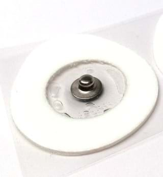
- 6 EMG sticker patch electrodes for large muscles
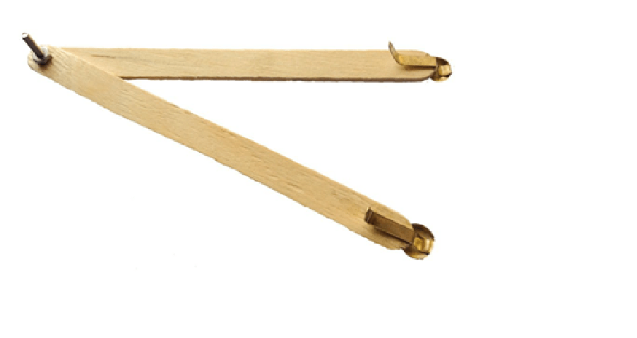
- 1 adjustable wooden electrode holder for small muscles
- 1 small bottle of electrode gel to be used with the small muscle electrode
- 1 smartphone cable to see/record spikes on mobile devices
- 1 laptop cable to view/record data onto a laptop and attach an external amplifier/speaker
- 1 Getting Started Guide
- 1 box (100 units) of EMG sticker patch electrodes for large muscles
- Access to Muscle SpikerBox Basics YouTube video (1:24 minutes)
- Access to metronome app
Note 1: This kit is ideal for you to do a demonstration. Alternatively, the above kit can support student teams of two or three. The setup could include a couple of stations that can facilitate EMG data collection for the entire classroom.
Note 2: Once the EMG data is collected, it is available to be analyzed for teaching or student research purposes.
Worksheets and Attachments
Visit [www.teachengineering.org/activities/view/umo-2936-virtual-lab-neuroscience-microcontrollers-activity] to print or download.Pre-Req Knowledge
None, although it is highly recommended to complete the first activity of the unit, Mapping the Brain: Neurons, Synapses, and Movement activity.
Introduction/Motivation
Breathing, seeing, speaking, swallowing, hearing, digesting, moving, sitting, and getting rid of waste are important bodily functions that all use what? (Let students offer answers. Answer: muscles.) Muscles are everywhere in the human body.
So, what are muscles? Muscle is a soft tissue in animals and humans that is made up of long cells that can contract and produce motion. Some muscles help move our body, and some help our internal organs keep us alive. Muscles are made of thousands of small fibers woven together. There are more than 650 muscles in your body that we use constantly. Do you think they are all the same? (Answer: No!)
What is unique about muscles that control the heart vs. the arms? Muscles use a combination of voluntary actions you can control, such as moving your arms and eyes, and involuntary movements that automatically happen, such as breathing and your heart beating, to work with nearly all of your body’s systems and functions. There are three types of muscle tissue in your body:
- Skeletal: Works with bones, tendons, and ligaments to support weight and help move
- Cardiac: Helps the cardiovascular (heart) system pump blood (mechanical contraction and relaxation)
- Smooth: Involuntary actions (e.g., respiratory, digestive, urinary)
Let us now focus on something we do in the classroom a lot: use of our wrists and fingers. Let us think about the various situations in which we use our fingers and wrists. What can we produce? (Potential answers: Writing, typing, texting, communicating.)
Do you think we can move the arms, wrists, and fingers with the same agility? What do we observe when we compare these three gestures—roll, pitch, and yaw—between fingers, wrists, and arms? Do they all move the same?
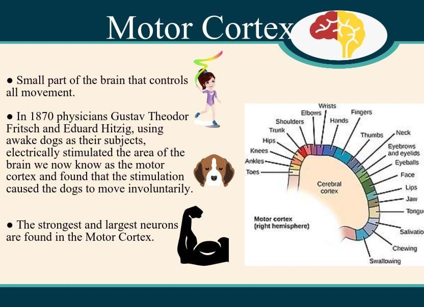
Now that you have made this observation, what do you think about how the brain communicates with the hands and legs? (Answer: Motor neurons that originate in the brain’s cerebral cortex, also known as the motor cortex, transmit electrical signals to the neuromuscular junction (NMJ) that cause muscles to contract. These finely tuned neural circuits are found throughout the body and innervate effector muscles and glands to enable both voluntary and involuntary motions.)
So, what do you think happens when our muscles have an issue? How can doctors identify what is going on?
In medicine, myopathy is a disease of the muscle in which the muscle fibers do not function properly. This meaning implies that the primary defect is within the muscle, as opposed to the nerves or elsewhere. If there are signs of muscle disease, nerve damage, or injury, an electromyography (EMG) test will help the doctor learn more about what is going on. An EMG test is often necessary to help diagnose or rule out conditions that include:
- Muscle disorders such as muscular dystrophy.
- Diseases affecting the connection between the nerve and the muscle.
- Disorders of nerves outside the spinal cord (peripheral nerves), such as carpal tunnel syndrome or peripheral neuropathies.
In this activity, we will investigate the neural origins of muscle movement based on data collected from the surface of an arm while performing simple hand gestures (e.g., wrist movement vs. finger movement). Our approach will use microcontrollers and skin-surface sticker electrodes to acquire EMG (muscle electrical signals) data for three gestures (roll, pitch, and yaw) at different frequencies. We will subsequently compare the difference in signals between large and finer muscle groups. Specifically, for this purpose, we will explore how the EMG signals differ when we do the same movement with our wrist compared to our fingers.
Procedure
Background
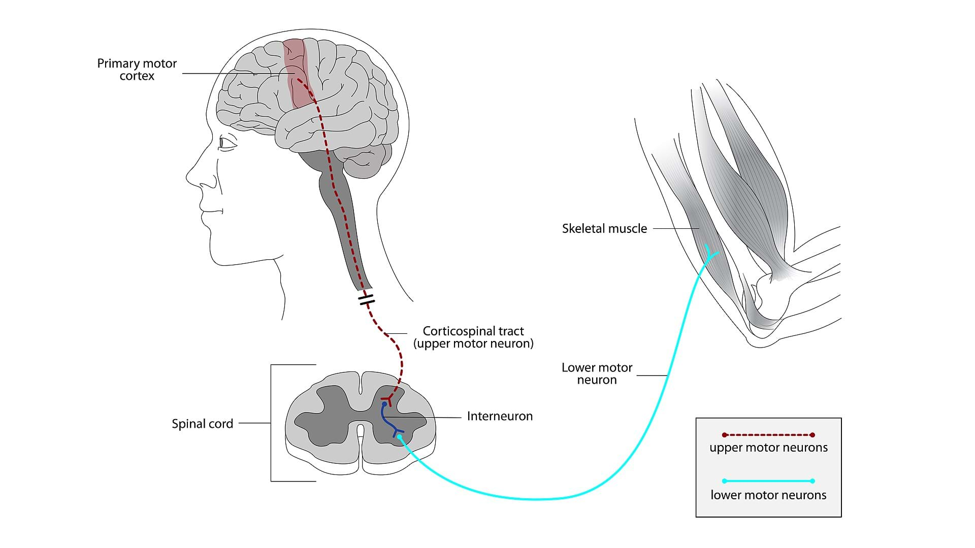
When your brain decides to move a muscle, electrical signals from your cerebral cortex (called "upper motor neurons") travel to your spinal cord, where they synapse with "lower motor neurons." These lower motor neurons then synapse with muscle to make a "motor unit."
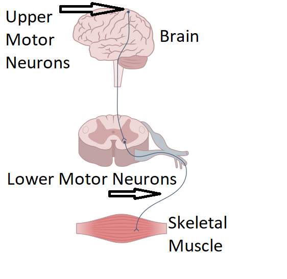
A single motor neuron can synapse with multiple muscle fibers. A motor unit is a single motor neuron and the multiple muscle fibers it innervates.
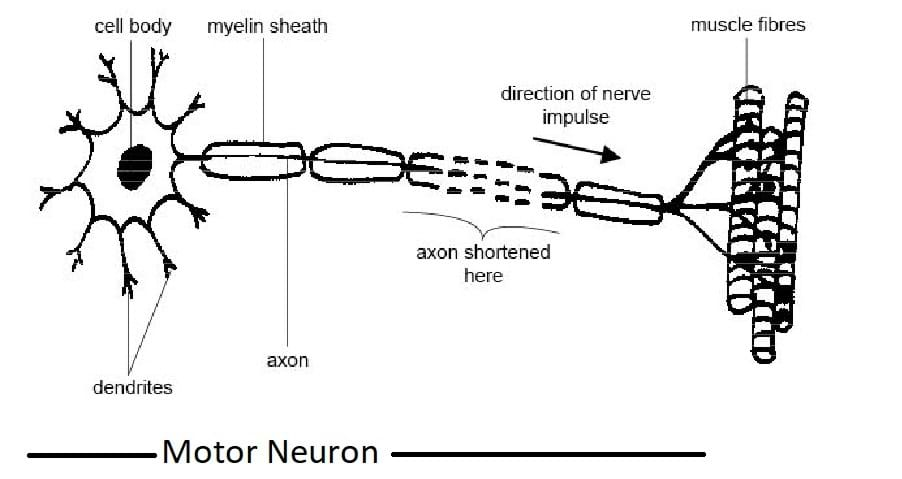
A muscle fiber is a very special type of cell that can change its shape due to myosin/actin chains sliding across each other. Specifically, muscle fibers are made up of blocks of proteins called myofibrils, which contain filaments that fold together when a muscle contracts. These fibers stretching and pressing together is what moves our organs or body. Myofibrils also contain a specialized protein (myoglobin), and molecules that provide the oxygen and energy needed for contraction.
In general, a large, powerful muscle like your bicep (or wrist) has motor neurons that innervate thousands of muscle fibers, whereas small muscles that require a lot of precision, such as your eyeball muscles or fingers, may have motor neurons that only innervate about 10 muscle fibers.
When a motor neuron fires an action potential, this causes a release of acetylcholine (ACh) at the synapse between the neuron and the muscle; this synapse is also called the neuromuscular junction. The acetylcholine then causes changes in the electrical potential of the muscle. Once this electrical potential reaches a threshold, an actual action potential occurs in the muscle fiber!
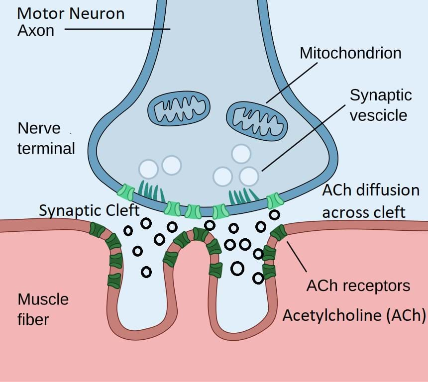
This action potential propagates across the membrane of the muscle, causing voltage-gated calcium channels to open, which begins the cellular cascade that causes muscle contraction.
When you contract a muscle, this is the result of many muscle fibers firing action potentials and changing shape. We can record this activity with the BYB Muscle SpikerBox.
The human wrist contributes to hand mobility and manipulation capabilities in healthy individuals. The wrist joint (also fingers) has three degrees of freedom (DOF) relative to the forearm:

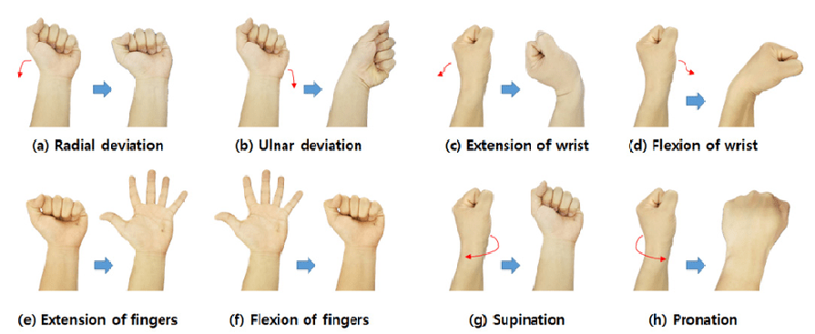
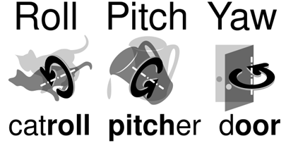
- Flexion-extension (Figure 6b (c and d)): Up and down movements (pitch)
- Pronation-supination (Figure 6b (g and h)): Rotational movements that occur at the joint (roll)
- Radial-ulnar deviation (Figure 6b (a and b)): Sideway movements (yaw)
What is an electromyogram? Electromyography (EMG) is a diagnostic procedure to assess the health of muscles and the nerve cells that control them (motor neurons). EMG results can reveal nerve dysfunction, muscle dysfunction, or problems with nerve-to-muscle signal transmission. An EMG uses tiny devices such as patch electrode stickers applied to the skin surface to measure the speed and strength of signals traveling between two or more points and translate these signals into graphs, sounds, or numerical values that are then interpreted by a specialist. Refer to the What is an EMG? Presentation for more details.
For this activity, students will measure wrist and finger movement signals over three dimensions of movement—roll, pitch, and yaw—that are naturally allowed. They will repeatedly test and record EMG data from the muscle vs. the finger before they analyze collected signals to recognize patterns. Using an inquiry-based learning approach, students will then begin to investigate neural origins of muscle movement and the differences in coarse (wrist) vs. finer movements (finger) based on data collected. Using a computational thinking approach, they will identify the various steps from the hardware processes, outlining the computational steps that enables the microcontroller, skin electrodes, and associated software to acquire EMG signals, process data, visualize, store, and analyze data.
The optimal experimental setup that will ensure repeatable, reliable, reproducible, and valid scientific data collection is as follows:
- Hardware:
- Laptop/Desktop
- BYB Muscle SpikerBox Bundle recommended for smooth data acquisition and analysis. This includes the following:
- Muscle SpikerBox
- 9V Battery (fully charged)
- EMG sticker patch electrodes (large muscles)
- adjustable wooden electrode holder (small muscles)
- electrode gel (for optimal contact with small muscles)
- orange electrode cable (3 alligator clips: 2 red, 1 black)
- laptop cable for data transfer
- Software:
- Spike Recorder software (PC/Mac/Linux version by Backyard Brains)
- Ensure software is correctly installed and responsive.
- Configure Spike Recorder to:
- Set Bandpass filter at 70-2500Hz.
- Enable 60Hz notch filter to reduce electrical noise.
- Electrode Placement:
- Large Muscle Electrodes (Wrist):
- Two large EMG sticker electrodes placed on opposite sides of the wrist (e.g., flexor/extensor surface).
- Reference electrode (black) attached away from recording site (e.g., upper arm, ring, or held in hand).
- Small Muscles Electrodes (Fingers):
- Adjustable wooden electrode holder used with small muscles in hand or finger area.
- Electrode gel applied for optimal signal quality.
- Experimental Procedure:
- Ensure proper electrode adhesion and connection; troubleshoot if necessary (e.g., verify battery charge, correct cable connections).
- Use an online metronome set to defined frequencies (40, 60, and 80 BPM).
- Clearly instruct participants to perform wrist and finger gestures (roll, pitch, and yaw) synchronized to metronome beats.
- Data collection duration: Two minutes per BPM setting per gesture type (wrist vs. finger movements).
- Clearly label and save recorded data as Wave files (.wav) in a dedicated experiment folder (e.g., "Wrist-40BPM-Yaw.wav").
- Data Acquisition and Validation:
- Confirm visual signal clarity using real-time visualization in Spike Recorder software.
- Practice the gestures first without recording, confirming clear transitions and identifiable EMG spikes.
- Validate signals before formal recording; troubleshoot noise issues by disconnecting charging cables, repositioning electrodes, or adjusting filters.
- Analysis and Interpretation:
- Compare recorded EMG data visually and numerically for differences in amplitude, frequency, and patterns between large and small muscle groups.
- Interpret differences between wrist and finger EMG signals, focusing on identifying characteristic signal patterns unique to specific movements or muscle groups.
- Troubleshooting Checklist for Optimal Performance:
- Electrical noise: Unplug chargers, reposition electrodes.
- Signal clarity: Confirm battery charge, electrode contact, cable integrity.
- Software responsiveness: Test Spike Recorder functionality before starting (e.g., microphone test—clap/snapping near the laptop).
- The above steps will ensure:
- Hardware integrity (battery power, correct electrode usage).
- Software optimization (appropriate filtering for clean signals).
- Experimental procedural guidelines (standardized metronome settings, gesture protocols).
- Troubleshooting steps to ensure rapid identification and correction of issues.
Before the Activity
- Gather BYB Muscle SpikerBox materials.
- Read the EMG Muscle SpikerBox Getting Started Guide.
- Follow the instructions in the Spike Recorder Documentation V2.0 to connect the Muscle SpikerBox to the computer.
- Download and install the BYB SpikerBox application to computer.
- Get familiar with setting up the BYB Muscle SpikerBox by reading Getting Started With Muscle SpikerBox.
- Make copies of the Muscle SpikerBox Pre-Assessment.
- Make copies of the Muscle SpikerBox Post Assessment.
- Make copies of the Muscle SpikerBox Project Report Format.
During the Activity
Day 1: Introduction and pre-activity assessment (50 min)
- Hand out one Muscle SpikerBox Pre-Assessment to each student.
- Give students 20 minutes to work on the assessment.
- Go through the Introduction and Motivation section and then the Background section of the activity.
- Have a class discussion about students’ answers to the Muscle SpikerBox Pre-Assessment.
- Answer any questions students may have.
Day 2: Introduction to Muscle SpikerBox Bundle (50 min)
- Show the following videos:
- Neuromuscular junction (9:00 minutes): https://www.khanacademy.org/science/health-and-medicine/human-anatomy-and-physiology/introduction-to-muscles/v/neuromuscular-junction
- Homunculus (5:15 minutes): https://www.khanacademy.org/science/health-and-medicine/nervous-system-and-sensory-infor/somatosensation-topic/v/somatosensory-homunculus
- Introduce students to the Backyard Brains (BYB) Muscle SpikerBox Bundle. Specifically, show students the Muscle SpikerBox, 9v battery, orange muscle electrode cable with three alligator clips (red, red, black), EMG sticker patch electrodes for large muscles, adjustable wooden electrode holder for small muscles, the cable to see/record spikes, and electrode gel.
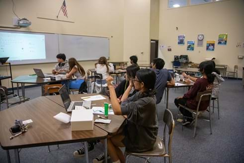
- Play the TED talk video (5:52 minutes) at https://youtu.be/rSQNi5sAwuc?feature=shared to explain the importance and potential of the Muscle SpikerBox equipment and how it can be associated with neuroscience.
- Divide the class into groups of three or four students.
- Instruct students to collect laptops and/or then download and install the BYB SpikerBox application from https://backyardbrains.com/products/spikerecorder.
Day 3: Set up and test the Muscle SpikerBox Bundle (50 min)
- Share the BYB pdf with the class: https://backyardbrains.com/products/files/SpikeRecorderDocumentation.2018.02.pdf
- Use this document to discuss and work with students to do the following:
- Connect the BYB SpikerBox to the computer.
- Configure inputs.
- Visualize data.
- Record data.
- Introduce students to the activity challenge:
- In this activity, you will explore how your wrist and finger muscles create movement in three dimensions—roll (rotation), pitch (up and down), and yaw (side to side). You will use sensors to repeatedly measure and record EMG signals from your muscles during different finger and wrist gestures. After collecting the data, you will analyze it to recognize patterns and compare larger, coarser wrist movements to finer finger movements.
- You will also take an inquiry-based approach to investigate where these movements originate in the nervous system and how signals travel from your brain to your muscles. Using computational thinking, you will break down the steps that make this all possible—from how the microcontroller and skin electrodes collect signals, to how software processes, visualizes, stores, and helps you analyze the data. This activity will give you a hands-on look at how biology, engineering, and coding all work together to help us understand the human body.
- Have students test the experimental setup (see Figures 3 and 4) to ensure hardware, software and electrodes work as expected. (Note: This is an important step to ensure data will be collected reliably.)
- Troubleshoot as necessary. Refer to the Troubleshooting Tips and Potential Safety Issues section to help students get set up.
- Once students are set up, have students try to clench their fist, move their wrist, and move their fingers to observe how the signals look in the software.
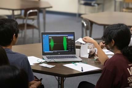
- Have students become familiar with data recording using the BYB Spike Recorder application.
Day 4: Introduction to EMG and experimental design (50 min)
- Optional: Review and reiterate the background information by drawing and/or showing students Figures 3, 4, and 5.
- Show the What is an EMG? Presentation and discuss what EMG is.
- Discuss muscle disorders/myopathies (e.g., Parkinson’s or carpal tunnel syndrome). Encourage students to see real-world connections and motivate their inquiry to apply critical thinking, scientific inquiry, engineering design principles, and creativity in a real-world context based on the EMG activities they performed.
- Example teacher script: “We have learned how muscles contract because of signals sent from the brain through motor neurons. But what happens if something goes wrong in this communication between nerves and muscles?
For instance, in Parkinson’s disease, certain nerve cells in the brain slowly break down. These nerve cells normally produce dopamine, a chemical messenger important for smooth and controlled muscle movements. When dopamine levels decrease, movements can become shaky (tremors), slow, stiff, or difficult to start. Parkinson’s is therefore a condition where communication between the brain and muscles is impaired, resulting in abnormal muscle activity visible in EMG recordings.
Another common issue is carpal tunnel syndrome, which involves pressure on the nerve in your wrist. When this nerve is compressed, signals between your brain and fingers slow down, causing symptoms such as pain, numbness, tingling, or weakness in the hand. EMG tests in this case often show delayed or weaker electrical signals traveling through the affected area, helping doctors diagnose the problem.
By studying EMG signals, like we are doing now, doctors and scientists can better understand these disorders, diagnose them earlier, and even design improved treatments. You might consider later how you could design experiments using EMG signals to better understand or even help treat these conditions.”
- Show Figures 6 A, B, and C and discuss specific activity (roll, pitch, and yaw in wrist vs. finger).
- Ask students to discuss placement of EMG electrodes based on Figures 6 A, B, and C. (Note: Students should review and discuss electrode placement strategies and start thinking about how these design considerations relate to identifying different types of movements or disorders. Here, students first begin hypothesizing how different experimental setups might yield different insights into neuromuscular pathologies or robotic applications.)
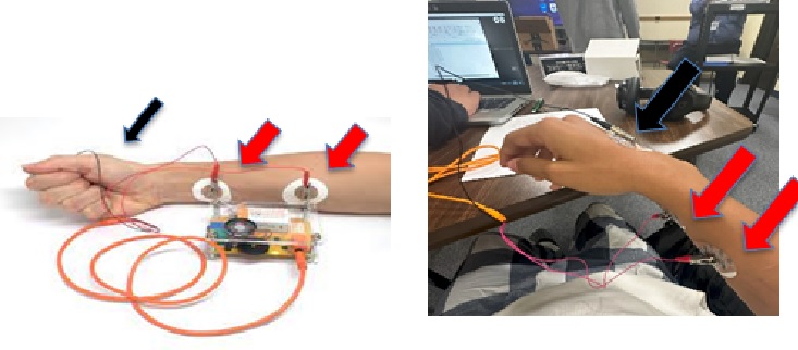
- Give students time to explore the BYB equipment by exploring hardware and software.
- Have students repeat Day 3 to become familiar with the BYB equipment and explore how the positioning of EMG electrodes impact signal data. They should try both EMG electrodes (Figures 7 a and b):
- EMG sticker patch electrodes for large muscles (wrist)
- Adjustable wooden electrode holder for small muscles (fingers)
Day 5: Experimental Procedure and Data Collection (50 min)
- Optional: Reiterate the activity challenge: In this activity, you will explore how wrist and finger muscles create movement in three dimensions—roll, pitch, and yaw—by measuring EMG signals and analyzing patterns in coarse and fine motor control. You will also investigate how these movements originate in the nervous system, using computational thinking to understand how biology, engineering, and coding work together to collect, visualize, and analyze data.
- Pass out or provide digital access to https://www.amnh.org/explore/videos/the-scientific-process.
- Before data collection, have students explicitly record their hypotheses about what they expect EMG signals to look like for different gestures and beats per minute (BPM) rates (using the provided Muscle SpikerBox Project Report Format to record these predictions.). This structured hypothesis formation will help them interpret testing results later and will help them form the basis for considering follow-up experiments.
- Divide the class into groups of three or four students.
- Describe how the students will work together: one person will record data, another person can ensure protocol is followed, one person is the subject, etc.
- Hand out one EMG Muscle Spike Box Getting Started Guide to each group.
- Have students open the downloaded and installed BYB SpikerBox application (if already downloaded to their computer or laptop). If needed, they can go to https://backyardbrains.com/products/spikerecorder to download the application.
- Have students gather their BYB Muscle SpikerBox materials as outlined in the EMG Muscle Spike Box Getting Started Guide.

- Give each group one Spike Recorder Documentation V2.0 handout.
- Have students follow the instructions in the Spike Recorder Documentation V2.0 handout to connect the Muscle SpikerBox to their computer.
- Have students test and troubleshoot if needed. Follow steps to ensure all connections are secure.
- Identify and discuss where students will place the electrodes for optimal recordings.
- Have students carefully place the large electrodes and then start recording EMG signals.
- Instruct students to save their data when they are finished with a particular BPM (e.g., they should name it by clearly naming files to identify wrist or finger data and BPM.)
- Have students proceed to the next BPM and repeat the protocol.
- Once each group has finished recording all BPM’s (40, 60, and 80) for the wrist, have them repeat the protocol for the finger.
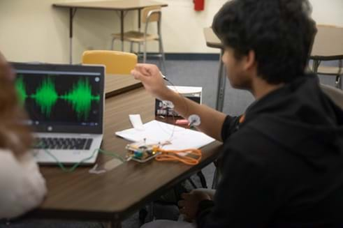
- Once the signals are recorded, have groups visualize using the SpikerBox application as outlined in the Spike Recorder Documentation V2.0 handout.
- Have groups discuss their findings.
- Have each group report their findings to the class.
Day 6: Data collection (50 min)
- Using the Day 5 instructions, give students the class period to collect data for fingers/wrist.
- Ensure that all data is reliably collected, correctly named, and saved.
Day 7: Post Assessment (50 min)
- Distribute the Muscle SpikerBox Post Assessment.
- Give students time to complete the Muscle SpikerBox Post Assessment.
- Lead a class discussion with the following questions. (Potential answers can be found in the Discussion Questions and Answers.)
- What were some limitations of the experimental design?
- What caused the spikes that you saw? Where did it originate? What do we know about the neuromuscular junction?
- What is occurring when the spike is positive? What is occurring when the spike is negative?
- If we were to redo the experiment, would you change the placement of electrodes? Where would be the best place to record from? Why?
- Identify the ways in which the experiment could be improved so that the results are consistently reliable, repeatable, and reproducible.
- How would the recordings differ if you were recording right inside the cell? What would be different about such an experimental setup? Would you see the same number and type of spikes? How would the amplitude change?
- What does muscle fatigue look like, how could you measure it, and what is causing this?
- Do different people have different rates of fatigue? Different muscles?
- Students will record their hypothesis and qualitatively compare the spike signals from wrist vs. finger movement.
- As a class, discuss, hypothesize, and suggest other experiments based on what they learned from these EMG studies (e.g., carpal tunnel syndrome, Parkinson’s, robotic/neuroprosthetic applications). Students may reflect on their observations, discussing considerations such as:
- Signal Differences: The EMG signals from finger muscles had lower amplitude but higher frequency spikes compared to wrist muscles. This may be due to smaller motor units and greater precision. Students could discuss exploring electrodes placed differently or using more precise sensors to see if signal quality and clarity improve, leading to better discrimination between gestures.
- Muscle Fatigue: The amplitude of EMG signals decreased over time, indicating muscle fatigue. Hence, muscle fatigue may correlate with reduced EMG amplitude and decreased frequency because fewer muscle fibers fire action potentials as they exhaust their energy sources. The wrist muscles fatigued more slowly than finger muscles because they’re larger and stronger. Students could design a prolonged exercise protocol to measure EMG changes during extended use, revealing patterns of muscular fatigue and recovery.
- Movement Complexity: Yaw movements had clearer and stronger EMG signals than roll or pitch movements, possibly because different muscles and degrees of freedom involve varying numbers of motor units.
- Sources of Error: Students noticed noise or spikes even when muscles were not moving, suggesting external interference or electrode placement issues.
- Neuromuscular pathologies: Students could propose follow-up investigations or alternative experimental designs. For example, they could propose to record EMG signals from patients associated with unusual muscle activation patterns, potentially aiding early detection or understanding progression of the disease. For example:
- Carpal tunnel investigation: If someone has carpal tunnel syndrome, the EMG signals from their fingers would be weaker or delayed compared to a healthy individual, as nerve conduction slows down.
- Parkinson’s disease EMG analysis: People with Parkinson’s disease might produce irregular EMG patterns, reflecting disrupted motor neuron firing patterns.
- Another topic of discussion could involve robotic, neurofeedback, and neuroprosthetic applications.
- Students could propose to use recorded EMG signals to control a robotic hand or arm, translating human muscle signals into precise movements. Testing accuracy and latency of different gestures could help improve prosthetic devices. By analyzing signal patterns in greater detail (e.g., using machine learning or Fourier analysis), students could consider designing a computer program that recognizes hand gestures purely from EMG data, enabling hands-free computer control or assistive technologies.
- Overall, examples such as those above will illustrate students’ ability to apply critical thinking, scientific inquiry, engineering design principles, and creativity in a real-world context based on the EMG activities they performed.
Day 8: Project Report (50 min)
- Hand out one Muscle SpikerBox Project Report Format to each group.
- Give students the rest of the period to complete their project report.
- Optional: If students run out of time, have them finish their reports as homework.
Vocabulary/Definitions
abduction: Moving a body part away from the center of the body, such as lifting an arm out and away from the body.
acetylcholine: Acetylcholine (ACh) is a neurotransmitter that motor neurons in the nervous system release at neuromuscular junctions to activate muscles and trigger voluntary movements.
adduction: Moving a body part toward the center of the body, such as bringing an arm back in so it rests along the torso.
axon: The portion of a nerve cell (neuron) that carries nerve impulses away from the cell body. A neuron typically has one axon that connects it with other neurons or with muscle or gland cells.
bandpass filter: An electronic circuit that allows a specific range of frequencies to pass through while attenuating frequencies outside that range.
beats per minute (BPM): The number of times a sound pulse beats in a minute.
corticospinal tract: A large bundle of axons that connects the cerebral cortex to the spinal cord to enable voluntary movement.
dendrite: A short, branched extension of a nerve cell, along which impulses received from other cells at synapses are transmitted to the cell body.
electrical noise: A disturbance in an electrical signal that can be unwanted or random, can be temporary or permanent, and can occur in both power and signal circuits.
electrode: An electrical conductor used to make contact with a nonmetallic part of a circuit. Electrodes carry electrical signals from muscles, brain, heart, skin, or other body parts to recording devices to help diagnose certain conditions.
extension: This movement increases the space between two body parts. An example of this is straightening the elbow.
flexion: A movement that brings two body parts closer together, such as the forearm and the upper arm.
hardware: A collective term used to describe any of the physical components of an analog or digital computer.
interneuron: A type of neuron that act as an intermediary between sensory and motor neurons in the central nervous system. They are the most common type of neuron in the body, making up over 99% of all neurons.
lower motor neuron (LMN): Those cells whose axons synapse directly on skeletal muscle. LMNs are located in the spinal cord and brainstem. The final muscle contraction is the product of all the interactions that have occurred in the central nervous system.
metronome: A practice tool that produces a steady pulse (or beat). The pulses are measured in beats per minute.
microcontroller: A compact integrated circuit designed to govern a specific operation in an embedded system; typically includes a processor, memory, and input/output (I/O) peripherals on a single chip.
mitochondria: An organelle (small structures) found in the cytoplasm (fluid that surrounds the cell nucleus) of a cell. Mitochondria make most of the energy for the cell and have their own genetic material that is different from the genetic material found in the nucleus.
muscle: A soft tissue in the body that is made up of thousands of elastic fibers that contract and relax to allow for movement. Muscles are attached to bones at either end and can move or stop the movement of a body part.
myelin sheath: An insulating layer, or sheath, that forms around nerves, including those in the brain and spinal cord. The myelin sheath allows electrical impulses to transmit quickly and efficiently along the nerve cells.
neuromuscular Junction: A synaptic connection between a neuron and a muscle cell.
neuron: Nerve cells that send messages all over the body to allow everything from breathing to talking, eating, walking, and thinking.
neurotransmitter: Chemical messengers that allow neurons to communicate with each other and other cells throughout the body. They are released by a neuron and travel across a synapse to affect a target cell, which can be another neuron, muscle cell, or gland. Neurotransmitters can act locally, affecting a small region of a postsynaptic cell, or more diffusely.
notch filter: A filter that attenuates frequencies within a very narrow range while passing all other frequencies unaltered.
pronation: The wheel-like rotation of the wrist toward the opposite shoulder.
radial deviation: Motion of the wrist/ finger toward the radial side (i.e., toward the thumb).
skeletal muscle: A highly organized tissue composed of bundles of muscle fibers called myofibers. Each myofiber represents a muscle cell with its basic cellular unit, the sarcomere.
software: Programming code executed on a computer processor. Software consists of computer programs that instruct the execution of a computer.
supination: The wheel-like rotation of the wrist away the opposite shoulder.
synapse: The space between the end of a nerve cell and another cell. Nerve impulses are usually carried to the neighboring cell by chemicals called neurotransmitters, which are released by the nerve cell and are taken up by another cell on the other side of the synapse.
synaptic cleft: A gap (~0.02 micron wide) between the pre- and postsynaptic nerve cells. The small volume of the cleft allows neurotransmitter concentration to be raised and lowered rapidly. Also called synaptic gap.
ulnar deviation: Motion of the wrist/ finger away from the thumb side.
upper motor neuron (UMN): Located in the cerebral cortex and brainstem, they send electrical impulses to initiate and regulate movement. UMNs send signals to lower motor neurons (LMN).
Assessment
Pre-Activity Assessment
Pre-Assessment: Students complete the Muscle SpikerBox Pre-Assessment.
Activity Embedded (Formative) Assessment
Project Report: Students complete the Muscle SpikerBox Project Report.
Post-Activity (Summative) Assessment
Post Assessment: Students complete the Muscle SpikerBox Post Assessment.
Post Activity Discussion: Students are prompted to answer these questions and then discuss as a group:
- What were some limitations of the experimental design?
- What caused the spikes that you saw? Where did it originate? What do we know about the neuromuscular junction?
- What is occurring when the spike is positive? What is occurring when the spike is negative?
- If we were to redo the experiment, would you change the placement of electrodes? Where would be the best place to record from? Why?
- Identify the ways in which the experiment could be improved so that the results are consistently reliable, repeatable and reproducible.
- How would the recordings differ if you were recording right inside the cell? What would be different about such an experimental setup? Would you see the same number and type of spikes? How would the amplitude change?
- What does muscle fatigue look like, how could you measure it, and what is causing this?
- Do different people have different rates of fatigue? Different muscles?
- Students will record their hypothesis and qualitati
Safety Issues
- Keep 9V batteries in original packaging until you are ready to use them. If loose, keep the 9V battery posts covered with masking, duct, or electrical tape. Prevent the posts from coming in contact with metal objects.
- Do not store 9V batteries loose in a drawer. Keep them someplace safe where they will not be tossed around. Do not store them in containers with other batteries.
Troubleshooting Tips
- If an error pops up during installation, check whether the Spike Recorder app is installed by checking for a shortcut on the desktop, going into the Apps and features menu, and checking the list of installed apps.
- Check the directory specified for the Spike Recorder app during installation. Make sure it is not in downloads.
- Within the Spike Recorder app, for EMG, set the bandpass filter to 70-2500 Hz, and tick the 60 Hz notch filter box.
- Also, if there is significant electrical noise observed while recording EMG signals, unplug the charger from both the laptop and the wall during the recording to reduce electrical noise.
- After connecting the Muscle Spike Recorder to the computer, start the Spike Recorder app. To ensure the app is responding, just clap or snap near the laptop near microphone to visualize sound data.
- Check that the cables are plugged in correctly and 9-volt (9V) battery has enough charge.
- If you cannot purchase the SpikerBox:
- As a low-cost alternative, you could use a micro:bit to obtain an analog signal and display that. Note: This is not the focus of this project, but it is possible. Given the limitations in signal processing, the dataset will be limited in scope, but as a proof of concept, a micro:bit could be used for signal acquisition in lieu of BYB Muscle SpikerBox.
- Because there are no identifiers on the attached data files, the .csv files could be downloaded and imported to continue on with the other activities.
Activity Scaling
For younger students/lower grades:
- Have a teacher demonstration and then a class discussion of EMG signals and the neuro-muscular circuit.
For advanced students/higher grades:
- Students can perform signal analysis (Fourier transform) and/or look up and apply different filters (notch vs. bandpass).
- Advanced students in bioengineering or engineering could explore the BYB SpikerBox circuit to learn about computer hardware.
- Advanced students in bioengineering could additionally explore how these signals could be interpreted and used as neuro-prosthetics to understand how neurorobotic arms work.
- Advanced students in computer science could write algorithms of how the SpikerBox application works and/or write code using Python to convert wave EMG signals to CSV files for plotting and analysis.
- Advanced students in medicine could explore neuromuscular or muscle-related pathologies and design experiments to explore them (e.g., Parkinson’s, carpal tunnel, age-related muscular dystrophy etc.)
Subscribe
Get the inside scoop on all things Teach Engineering such as new site features, curriculum updates, video releases, and more by signing up for our newsletter!More Curriculum Like This

Students learn about the function and components of the human nervous system, which helps them understand the purpose of our brains, spinal cords, nerves and five senses. In addition, how the nervous system is affected during spaceflight is also discussed.

This is the first activity in a unit of four activities. Students are introduced to foundational neuroscience concepts, focusing on motor and sensory neurons and their connections to muscles at the synapse. Through handouts and activities, they explore brain anatomy and construct a basic homunculus ...

Students learn about the similarities between the human brain and its engineering counterpart, the computer. Since students work with computers routinely, this comparison strengthens their understanding of both how the brain works and how it parallels that of a computer.

Students learn about human reflexes, how our bodies react to stimuli and how some body reactions and movements are controlled automatically, without thinking consciously about the movement or responses. In the associated activity, students explore how reflexes work in the human body by observing an ...
References
Byun, Sung-Woo, and Seok-Pil Lee. “Implementation of Hand Gesture Recognition Device Applicable to Smart Watch Based on Flexible Epidermal Tactile Sensor Array.” Micromachines vol. 10,10 692. 12 Oct. 2019, doi:10.3390/mi10100692.
Cleveland Clinic. “Muscle.” Cleveland Clinic, 29 Sept. 2021,my.clevelandclinic.org/health/body/21887-muscle.https://my.clevelandclinic.org/health/body/21887-muscle.
Dobrowolny, Gabriella et al. “Age-Related Alterations at Neuromuscular Junction: Role of Oxidative Stress and Epigenetic Modifications.” Cells vol. 10,6 1307. 24 May. 2021, doi:10.3390/cells10061307.
Haggie, Lysea et al. “Linking cortex and contraction-Integrating models along the corticomuscular pathway.” Frontiers in physiology vol. 14 1095260. 10 May. 2023, doi:10.3389/fphys.2023.1095260.
Getting Started with Spike Recorder on PC/Mac/Linux How Do I Connect SpikerBox to Computer Using USB Cable? https://backyardbrains.com/products/files/SpikeRecorderDocumentation.2018.02.pdf.
“Lesson Explainer: Muscle Contraction | Nagwa,” www.nagwa.com/en/explainers/676192534258.
LibreTexts. “13.1: Muscle Contraction.” Biology LibreTexts, 7 Oct. 2016, bio.libretexts.org/Bookshelves/Introductory_and_General_Biology/Introductory_Biology_(CK-12)/13%3A_Human_Biology/13.01%3A_Muscle_Contraction, https://bio.libretexts.org/Bookshelves/Introductory_and_General_Biology/Introductory_Biology_(CK-12)/13%3A_Human_Biology/13.01%3A_Muscle_Contraction.
Mayo Clinic. “Electromyography (EMG) - Mayo Clinic.” Www.mayoclinic.org, 21 May 2019, www.mayoclinic.org/tests-procedures/emg/about/pac-20393913#:~:text=Electromyography%20(EMG)%20is%20a%20diagnostic. https://www.mayoclinic.org/tests-procedures/emg/about/pac-20393913#:~:text=Electromyography%20(EMG)%20is%20a%20diagnostic,%2Dto%2Dmuscle%20signal%20transmission.
“Neuromuscular Junction, Motor End-Plate (Video).” Khan Academy, www.khanacademy.org/science/health-and-medicine/human-anatomy-and-physiology/introduction-to-muscles/v/neuromuscular-junction.
Palmer, A K et al. “Functional wrist motion: a biomechanical study.” The Journal of hand surgery vol. 10,1 (1985): 39-46. doi:10.1016/s0363-5023(85)80246-x,
http://usdbiology.com/cliff/Courses/Behavioral%20Neuroscience/CStart/2%20Fundamentals%20of%20Neurocircuitry%20III.html.
Sato, Yoichi et al. “Real-time input of 3D pose and gestures of a user's hand and its applications for HCI.” Proceedings IEEE Virtual Reality 2001 (2001): 79-86.
“Somatosensory Homunculus.” Khan Academy, 2023, www.khanacademy.org/science/health-and-medicine/nervous-system-and-sensory-infor/somatosensation-topic/v/somatosensory-homunculus
Copyright
© 2025 by Regents of the University of Colorado; original © 2024 University of MissouriContributors
Dr. Ashwin Mohan, computer science faculty at the Illinois Mathematics and Science Academy (IMSA); Dr. Sowmya Anjur, emeritus science faculty from IMSA; Mrs. Namrata Pandya, retired computer science faculty from IMSA; Matthew Stroud, pre-med student at the University of Missouri; Dr. David Bergin, emeritus, College of Education & Human Development, University of Missouri; and Dr. Satish Nair, professor of electrical engineering and computer science at the University of Missouri.Supporting Program
Research Experience for Teachers (RET), University of Missouri ColumbiaAcknowledgements
This work is based on work supported in part by the National Science Foundation under grant no. EEC-1801666—Research Experiences for Teachers at the University of Missouri. Any opinions, findings and conclusions or recommendations expressed in this material are those of the authors and do not necessarily reflect the views of the National Science Foundation.
This endeavor would not have been possible without the guidance and support of Prof. Satish S. Nair, University of Missouri, Neural Engineering Laboratory, Columbia, Missouri (https://nairs.mufaculty.umsystem.edu/home).
We wish to thank the Illinois Mathematics and Science Academy (IMSA), Aurora, IL, where student testing was conducted.
Last modified: July 3, 2025





User Comments & Tips