Quick Look
Grade Level: 11 (9-12)
Time Required: 45 minutes
Expendable Cost/Group: US $15.00
Group Size: 3
Activity Dependency:
Subject Areas: Biology, Life Science, Science and Technology

Summary
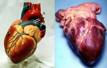
Students learn about the form and function of the human heart through the dissection of sheep hearts. They learn about the different parts of the heart and are able to identify the anatomical structures and compare them to the all of the structural components of the human heart they learned about in the associated lesson.Engineering Connection
Engineers are very involved in designing and testing new materials to improve quality of life and prolong human life expectancy. For example, biomedical engineers develop and test new materials to replace faulty heart valves. Biomedical engineers who design technologies to help patients with cardiovascular diseases must understand the structure and functions of the heart, including how blood flows through the heart, and the stresses that this imparts on heart tissue.
Learning Objectives
After this lesson, students should be able to:
- Identify the parts of the human heart on a diagram and with biological specimens, specifically, the left and right ventricles, left and right atria, interventricular septum, the mitral, tricuspid, pulmonary, and aortic valves, pericardium (if present), valve leaflets, and aorta.
- Describe blood flow through the human heart, elaborating on what role each part of the heart plays in this process.
- Define key terms associated with the heart and its function, including the endocardium, myocardium, regurgitation, pericardium, valve, blood flow.
Educational Standards
Each Teach Engineering lesson or activity is correlated to one or more K-12 science,
technology, engineering or math (STEM) educational standards.
All 100,000+ K-12 STEM standards covered in Teach Engineering are collected, maintained and packaged by the Achievement Standards Network (ASN),
a project of D2L (www.achievementstandards.org).
In the ASN, standards are hierarchically structured: first by source; e.g., by state; within source by type; e.g., science or mathematics;
within type by subtype, then by grade, etc.
Each Teach Engineering lesson or activity is correlated to one or more K-12 science, technology, engineering or math (STEM) educational standards.
All 100,000+ K-12 STEM standards covered in Teach Engineering are collected, maintained and packaged by the Achievement Standards Network (ASN), a project of D2L (www.achievementstandards.org).
In the ASN, standards are hierarchically structured: first by source; e.g., by state; within source by type; e.g., science or mathematics; within type by subtype, then by grade, etc.
International Technology and Engineering Educators Association - Technology
-
Assess how similarities and differences among scientific, mathematical, engineering, and technological knowledge and skills contributed to the design of a product or system.
(Grades
9 -
12)
More Details
Do you agree with this alignment?
State Standards
Tennessee - Science
-
Explore the anatomy of the heart and describe the pathway of blood through this organ.
(Grades
9 -
12)
More Details
Do you agree with this alignment?
-
Describe the biochemical and physiological nature of heart function.
(Grades
9 -
12)
More Details
Do you agree with this alignment?
Materials List
Each group needs:
- 1 sheep heart, for $4.75-6.00 each at www.carolina.com
- dissection kit (scalpel, pins, probe, scissors), for $24 at https://www.amazon.com, generally ranging from $10-30, or a set of 10 for $60
- dissection tray (if the dissection kit does not include a tray) for $10-20 at https://www.amazon.com; alternative, a basic cake pan
- protective gear for each student
- aprons, one per student
- disposable gloves, one per student
- lab goggles, one per student
- access to a clutter-free desk or lab bench
- vinyl tablecloth (to cover the desk or lab bench)
- 6-7 sheets of newspaper (to lay beneath the vinyl tablecloth to further protect the surface); alternative: poster/butcher block paper
- small kitchen trash bag
To share with the entire class:
- (optional) pictures of previous dissections
- paper towels
- cleaning materials for the lab
- 1-2 50-gallon lawn and leaf/trash bags
Worksheets and Attachments
Visit [www.teachengineering.org/activities/view/van_heartvalves_lesson01_activity1] to print or download.Pre-Req Knowledge
It benefits students to already know how blood flows through the heart, as covered in the associated lesson of this unit, Heart to Heart, or the What Do I Need to Know about Heart Valves? lesson.
Introduction/Motivation
The human heart is vital in sustaining homeostasis, which means it helps to regulate the internal environment of our body. This includes conditions such as internal temperature and pH. Our hearts have a specific anatomy that aids in this function. The way in which the heart is designed determines the path that blood must take in order to be pumped around the body. Without this awesome design, our hearts would not be able to supply every single cell in our bodies with vital oxygen and nutrients.
Ever wonder how many cells are in a human body? Although an exact average is not known, it is believed that a human body has around 50 trillion cells. Each cell needs oxygen and nutrients, as well as a way to get rid of waste, including carbon dioxide. Does anyone know how our bodies are capable of taking care of the needs of 50 trillion cells? Believe it or not, our blood is the answer to this question! Our blood carries nutrients, hormones, and of course, oxygen, throughout the body to provide cells with everything they need to maintain body temperature, pH, and even to fight diseases and infections.
Now that we know as many as 50 trillion cells need to be reached by our blood, we can appreciate the importance of the design of the human heart. It is an amazing muscular machine! Today, you are going to dissect a sheep heart and learn exactly what makes a heart so well designed and mighty and how it gives us life every day.
Procedure
Background
For the heart dissection, have students work in groups of two or three. Have the dissecting materials available prior to the start of the lab, and review safety precautions with the class. For the dissection, have students gather their supplies and follow instructions from the teacher. At the end of the dissection, have students dispose of all specimens and disposable material safely, and clean up and disinfect all tools and surfaces. Check with your school board for biological specimen disposal procedures, which can range from discarding in the classroom trash bins (double-bagged) to arranging for a hazardous waste disposal company's services.
Before the Activity
- Rinse each sheep heart with cold water and lay it on a paper towel or newspaper to drain and dry.
- Make copies of the Heart Dissection Protocol, one for each student.
With the Students
Part I: Dissection
- Distribute the protocol handouts. This handout includes the instructions listed below. If students miss any of the spoken instructions, they will have the written protocol to follow and reference.
- Inform students that they will be drawing the heart from each angle that they observe it from and labeling its structures. (Note: all of the words shown in bold in the steps below are structures that students should identify, draw and label. Make sure to circulate the room and check that students are identifying each structure correctly at each step.)
- If any sheep heart has a pericardium intact, tell students to observe it before removal. Students may view another group's heart if their specimen is not intact.
- Instruct students to observe the external anatomy of the heart. Find the coronary arteries (they may need to scrape away a little bit of the observed fat to find the coronary arteries). Also have them note the two flaps that are located on the top of the heart. These are the auricles, which are part of the atria of the heart.
- Have students note the vessels that are leaving the top of the heart. These are the pulmonary trunk, which leads to the pulmonary arteries, and the arch of the aorta. To differentiate between the two, the pulmonary truck is the more anterior of the two. Tell students to cut into the pulmonary trunk and the aorta, making note of the valves they see there. These are semilunar valves, with the aortic valve being in the aorta, and the pulmonary valve being in the pulmonary trunk.
- Direct students to hold the heart so that they can observe the posterior (opposite side). Look for the four pulmonary veins that enter into the left atrium (these vessels may not be present, so direct students to use their probes to find the openings where they would have been located). Next, look for the superior and inferior vena cavae entering the right atrium.
- Use the scissors provided in the dissection kit to cut through the wall of the inferior vena cava and into the right atrium. Note the valve at the bottom of the atrium. This is the tricuspid valve.
- Using the dissection kit scalpel, cut from the right atrium downward through the myocardium. You are now cutting into the right ventricle. Using a probe, follow the right ventricle into the pulmonary artery.
- Use the scalpel again to cut from the left atrium downward. At the bottom of the left atrium, you should be able to see the mitral (bicuspid) valve. Continuing down, you should see the left ventricle. Note how thick the myocardium of the left ventricle is compared to the right ventricle. Use a probe to follow the left atrium up into the aorta.
Part II: Clean Up
- While still wearing gloves, have students carefully lay scissors, scalpels and probes on a separate piece of newspaper.
- Ask them to carefully lift the sides of their vinyl tablecloths such that they gather all of their sheep heart parts into the center. Wrap up the contents tightly and put them into a kitchen trash bag.
- Put the kitchen trash bag into a large 50-gallon trash bag. Remove the disposable gloves and drop them into the large trash bag.
- Leave the dissection tools (scissors, scalpels and probes) and goggles on the newspaper for the teacher. After disinfection, the dissection tools and goggles can be reused.
Vocabulary/Definitions
aorta: The largest artery of the body, which takes blood from the left ventricle and moves it to the body.
atria: The two chambers of the heart that receive blood from the body.
endocardium: The innermost layer of the heart that touches blood that is flowing through the heart
epicardium: A tissue layer that covers the myocardium of the heart and makes up the outside borders of the heart.
homeostasis: A process in which a body maintains and regulates its internal environment, including pH and temperature.
myocardium: Thick muscle tissue that makes up the middle layer of the heart.
pericardium: A membrane that covers and protects the heart.
pulmonary circulation: Movement of blood between the heart and the lungs.
systemic circulation: Movement of blood between the heart and the body (excluding the lungs).
valve: A structure (of the heart) that controls the flow of a fluid, as one that permits blood to flow in one direction only.
ventricles: The two chambers of the heart that receive blood from the atria and send blood to the body.
Assessment
Pre-Activity Assessment
Why is this Important? Brainstorm and Sketch: Have students work in pairs, or groups of three at most, to brainstorm and list all the reasons they believe the heart is important. Ask them to consider what jobs the heart must perform and how the heart is structured so that it functions as necessary. After brainstorming, instruct students to sketch a drawing of what they think a human heart looks like. As best as possible, have them label the chambers and different structural parts of the heart. Ask a few student volunteers share their work with the class.
Activity Embedded Assessment
What Do You See? Sketch: Have students draw the sheep's heart in detail. Each student should sketch what s/he sees from multiple angles. Collectively, students should make sure their group's drawings include the following structures: left and right atria, left and right ventricle, tricuspid valve, mitral valve, interventricular septum, aorta, aortic valve, and the spaces where the superior and inferior vena cava, pulmonary arteries, and pulmonary veins enter the heart. Label each structure. If students have difficulty locating any of structure, encourage them to ask the teacher for assistance during the dissection.
Post-Activity Assessment
Heart Review Quiz: Once the activity is completed and cleaned up, show students an unlabeled diagram of the heart. Do this as a handout or projected overhead transparency or PowerPoint® slide. If the blank diagram is shown to the entire class at once, point out the various parts of the heart, including the following structures: left and right atria, left and right ventricle, tricuspid valve, mitral valve, interventricular septum, aorta, aortic valve, and the spaces where the superior and inferior vena cava, pulmonary arteries, and pulmonary veins enter the heart. Ask students to write down on a piece of paper the name of each structure. If the diagram is provided as a handout, have students label each structure. Collect their answers and grade as a quiz.
Sources of unlabeled heart diagrams suitable for this quiz include: http://www.smartdraw.com/circulatory-system-diagram/examples/cardiac-circulation/ and http://www.smm.org/heart/lessons/heartDiagram.htm.
Another option is to use one of the sheep hearts instead of an unlabeled diagram. Wearing disposable gloves and holding up the heart in front of the entire class, point to each structure while students independently record on paper the names of the structures. Have students turn in their answers for grading. Review their answers to ascertain their level of comprehension.
Homework
Use Your Imagination: Ask students to design a new technology that could be created through engineering and is inspired by the heart. Tell them to consider the strength and power of the heart, as well as its ability to direct and move the blood. Encourage students to be as creative as possible. The technology does not have to be built, but they should include a drawing and an explanation of what their technology is, how it functions, and how it is inspired by the heart.
Safety Issues
- Make students aware of the location of an eyewash station, and the emergency procedures while conducting a lab.
- During dissection, remind students that they must wear the protective aprons, gloves and eyewear.
- Since dissections requires the use of sharp instruments, coach students on the safe use of scalpels and scissors. Account for all scalpels and scissors before students leave the room at the end of the lab period.
Troubleshooting Tips
During the dissection, closely monitor the groups to make sure they know what structures they are looking at, and make sure they are following safe practices.
Subscribe
Get the inside scoop on all things Teach Engineering such as new site features, curriculum updates, video releases, and more by signing up for our newsletter!More Curriculum Like This

Students learn about the form and function of the human heart through lecture, research and dissection. They brainstorm ideas that pertain to various heart conditions and organize these ideas into categories that help them research possible solutions.

Students learn how healthy human heart valves function and the different diseases that can affect heart valves. They also learn about devices and procedures that biomedical engineers have designed to help people with damaged or diseased heart valves.

This lesson describes how the circulatory system works, including the heart, blood vessels and blood. Students learn about the chambers and valves of the heart, the difference between veins and arteries, and the different components of blood.

Students learn all about the body's essential mighty organ, the heart, as well as the powerful blood vascular system. This includes information on the many different sizes and pervasiveness of capillaries, veins and arteries, and how they affect blood flow through the system. Then students focus on ...
References
Shier, D., Butler, J. and Lewis, R. Hole's Human Anatomy & Physiology, Eleventh Edition. New York, NY: McGraw Hill Higher Education, 2007.
Copyright
© 2013 by Regents of the University of Colorado; original © 2011 Vanderbilt UniversityContributors
Michael Duplessis; Janet Yowell; Carleigh SamsonSupporting Program
VU Bioengineering RET Program, School of Engineering, Vanderbilt UniversityAcknowledgements
The contents of this digital library curriculum were developed under National Science Foundation RET grant nos. 0338092 and 0742871. However, these contents do not necessarily represent the policies of the NSF, and you should not assume endorsement by the federal government.
Last modified: February 9, 2018


User Comments & Tips