
Heart to Heart
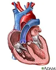 The human heart.Copyright U.S. National Library of Medicine, U.S. Department of Health and Human Services http://www.adamimages.com
Students learn about the form and function of the human heart through lecture, research and dissection. They brainstorm ideas that pertain to various heart conditions and organize these ideas into categories that help them research possible solutions. An expert in the field of cardiac valve research was interviewed for this lesson and shares his ideas with the class. Students conclude by researching various possible heart defects.
Engineers begin their work to design solutions to problems by first gathering information in the form of background information about the system they are working with, testing and analysis data that they collect from various sources, and information from experts on the subject matter.
After Part I of this lesson, students should be able to:
- Identify the parts of the heart (left and right ventricles, left and right atria, interventricular septum, mitral valve, tricuspid valve, pulmonary valve, aortic valve, pericardium, valve leaflets, aorta).
- Describe how blood flows through the heart in a specific path.
- Explain how problems with the heart may cause health concerns.
After Part II of this lesson, students should be able to:
- Identify the parts of the human heart on a diagram and with a biological specimen.
- Describe blood flow through the human heart, elaborating on what role each part of the heart plays in this process.
- Define terms associated with the heart and its function.
- Explain what blood pressure is, and take blood pressure using available tools.
- STEL-1O Assess how similarities and differences among scientific, mathematical, engineering, and technological knowledge and skills contributed to the design of a product or system.
Grades 9-12
Do you agree with this alignment?
- 3251.4.6 Describe the biochemical and physiological events associated with heart contraction, blood pressure, and blood clotting.
Grades 9-12
Do you agree with this alignment? - CLE 3251.4.2 Explore the anatomy of the heart and describe the pathway of blood through this organ.
Grades 9-12
Do you agree with this alignment? - CLE 3251.4.3 Describe the biochemical and physiological nature of heart function.
Grades 9-12
Do you agree with this alignment? - CLE 3295.T/E.4 Describe the dynamic interplay among science, technology, and engineering within living, earth-space, and physical systems.
Grades 9-12
Do you agree with this alignment?
Part I: Legacy Cycle
After presenting the "grandmother" challenge, hand out the Challenge Question Brainstorm Worksheet (PDF) and ask students to brainstorm ideas about what could be causing the heart condition (Legacy Cycle Step 2: Generate Ideas). Hopefully, the challenge information provided will steer students towards a possible cardiac issue, but be prepared for other ideas (such as respiratory issues, infection, etc.). Record all ideas on the classroom board, and group them into student-generated categories. While there is no real lecture to this part of the lesson, it is important to that all students participate in the brainstorming portion of the lesson and record all ideas. In brainstorming, remind students that no ideas are silly, and that all ideas should be heard. Hopefully, students have some early ideas they can share about the heart, but it may be necessary to prompt them if not.
After students have completed brainstorming ideas, read to them (or have them read to themselves) the Cardiac Valve Research: Expert Interview (PDF) (Legacy Cycle Step 3: Multiple Perspectives), which introduces ideas about how heart valves might contribute to health problems. This interview steers them more towards a cardiac issue and a solution. After reading the interview, begin a class discussion about heart defects. (Note: You may need to distinguish between congenital defects (present at birth) and those that develop later in life.)
A congenital heart defect is a condition that approximately 9 of every 1,000 people are born with and affects the structure of the heart and the large blood vessels that directly move blood to and from the heart (aorta, pulmonary arteries, superior and inferior vena cava). These defects can block blood flow or cause blood to flow in irregular patterns through the heart. In some cases, no action needs to be taken, but in other cases, medication or surgery is necessary.
While a congenital heart defect is a condition that some people are born with, many other heart diseases and defects develop through people's lives. These conditions, referred to as cardiovascular or heart disease, affect the heart's valves, muscles and blood vessels, including arteries, capillaries and veins. These diseases include coronary heart disease (which affects the coronary artery that supplies blood to the heart muscles, and can lead to heart attack)s, cardiomyophathy (a disease of the muscles in the heart), hypertensive heart disease (includes complications to the heart due to high blood pressure), valvular heart disease (diseases such as inflammation or stiffness to the heart valves that affects the movement of blood through the heart), and many others.
Part I of the lesson ends with students using the Internet to research possible heart problems by (Legacy Cycle Step 4: Research and Revise). They should research the diseases previously mentioned, as well as other heart diseases. Remind students that the goal is to search for information that might be useful in approaching the challenge question. (Note: If no time remains, assign completion of this research as homework.) Suggested search topics include heart valve disease, heart valve problems, cardiac regurgitation and heart function.
Part II: Heart Anatomy and Blood Flow
Part II of the lesson (Days 2-4) utilizes Legacy Cycle Step 4: Research and Revise in that students learn about the form and function of the heart, as well as about blood flow through the heart's chambers. This information is useful in later understanding the concept of blood pressure, as well as the stresses placed on heart valves and the problems caused by such stress. Students also research the anatomy of the heart by dissecting a sheep heart in the associated activity, The Mighty Heart.
On Day 2 of the lesson, provide a class lecture using The Human Heart and Blood Flow Presentation (PPTX) (a PowerPoint file) and the suggested script below, on heart anatomy and blood flow. The purpose of the lecture is to ensure students understand how the heart is structured and how it works; spending a quality amount of time on this background information ensures student mastery of heart form and function and lends clarity to the activities.
The information below is a suggested script for the PowerPoint presentation. While intended to map to the slides, it is also great resource information on its own, or in conjunction with detailed heart photographs or diagrams.
Slide 1: Title: The Human Heart and Blood Flow
Slide 2: The human heart is a muscular organ located in the thoracic cavity, nestled between the two lungs and very slightly to the left of center. For adult humans, it is about the size of their clenched fists; for children, it is about the size of their clenched fists, and grows as they grow. The heart weighs approximately ½ pound, but it is normal to vary in size, just as the overall size of people varies.
Slide 3: The heart, like many organs in the body, is covered by a protective membrane. This membrane is called the pericardium. The pericardium also holds fluid, which adds additional protection to the heart. This fluid is called pericardial fluid. The heart itself is composed of muscle tissue, and is subdivided into three layers:
- Epicardium — this is the outermost layer of the heart, and it helps to reduce friction by containing some fat amongst the coronary arteries.
- Myocardium — this thick layer of cardiac muscle is the part of the heart that contracts to create a pumping action. It is very thick around the left ventricle because of the amount of force needed to move the blood from the heart to the rest of the body.
- Endocardium — this is the innermost layer of the heart, and comes into physical contact with the blood. This layer lines the two atria and the ventricles.
Slide 4-5: The heart is a pump that is designed to move blood through the human body. It does this by contractions of the myocardium, thus causing the chambers of the heart to compress and push the blood within. Valves serve as one-way doors, separating the chambers of the heart and the vessels that lead out of the heart. These valves prevent blood from moving in the wrong direction as it moves through and out of the heart. Contractions occur at an average rate of 70 beats per minute (bpm), providing the movement of a continual flow of blood throughout the body.
Slides 6-7: Blood moving through the heart is cyclic, so we start with blood entering the heart after it has been through the rest of the body. We call this blood deoxygenated because the cells of the body have diffused the oxygen from the red blood cells for use in cellular respiration. Deoxygenated blood follows the following path:
- Superior and inferior vena cava — deoxygenated blood enters the heart from these two veins. The superior vena cava drains the upper portions of the body (head and arms), while the inferior vena cave drains the lower parts of the body. This blood enters the...
- Right atrium — deoxygenated blood enters the heart through this chamber. The coronary sinus is another smaller vein that drains the myocardium in to this chamber. When the atria contract, this pushes blood down through the...
- Tricuspid valve — a three-flap valve that opens whenever blood is pushed through it by the contractions of the atria. This valve also closes whenever the atria relax because of the change in pressure from the blood. To prevent the valves from folding back the incorrect way, they are held in place by long fibers called chordae tendineae.
- Right ventricle — this chamber receives deoxygenated blood from the right atrium, and contracts to push blood up to the lungs. This muscular chamber has thinner walls than the left ventricle simply because the distance the right ventricle has to pump blood is shorter. As it leaves the right ventricle, the blood passes through the...
- Pulmonary (semilunar) valve — this valve also has three leaflets (cusps) that close to prevent backflow (regurgitation) of blood. From here, blood travels into the...
- Pulmonary arteries — these arteries carry deoxygenated blood from the heart to the lungs. This is the only artery in the body to carry oxygen-poor blood, because it is on its way to the lungs to pick up more oxygen.
- Pulmonary veins — re-oxygenated blood reenters the heart via these veins, which are the only veins in the body to carry oxygen-rich blood. This blood reenters the heart through the...
- Left atrium — this chamber contracts with the right atrium, and pushes blood through the...
- Mitril (bicuspid) valve — this valve has two leaflets, and also has the chordae tendineae to reinforce them.
- Left ventricle — this chamber receives blood from the left ventricle, and is responsible for contracting to push blood out through the vessels of the body. Because of this task, the myocardium that makes up the walls of the left ventricle is much thicker. Also, the left and right ventricles are separated by a myocardial wall called the interventricular septum. Blood leaving this chamber moves through the...
- Aortic (semilunar) valve — this valve works much the same way as the pulmonary valve works.
- Aorta — this is the largest artery of the body, receiving oxygen-rich blood from the left ventricle and moving it through the rest of the body via other smaller arteries and veins.
Have students continue their research on congenital heart defects and the nature and side effects of valve disease for additional learning opportunities (or assign this work as homework). Many online resources are at students' disposal. A focused internet search using the keywords previously mentioned yields hundreds of content-rich documents.
Specifically, additional information on congenital heart defects can be found at: http://kidshealth.org/parent/medical/heart/congenital_heart_defects.html#
And, additional information on valve disease can be found at: http://www.medicinenet.com/heart_valve_disease/article.htm
Your grandmother, who has been a non-smoker and relatively healthy throughout her life, has recently noticed that she is becoming increasingly short of breath as she does simple things, such as climbing the steps in her house. She has also noticed that her heart rate increases when she does mild exercise, such as walking to the mailbox or going upstairs. What could be causing this? Is it just due to her getting older? Is there reason to be concerned? What can be done to help her?
The human heart is vital in sustaining homeostasis—the stability or equilibrium in a biological system. In fact, the heart has a specific anatomy that aids in this function. The way the heart is designed determines the path that blood must take through it in order to be pumped around the body.
- valve
- A flap of tissue that acts as a one-way door to help blood flow in one direction in the heart.
- myocardium
- Thick muscle tissue that makes up the middle layer of the heart.
- epicardium
- A tissue layer that covers the myocardium of the heart and makes up the outside borders of the heart.
- endocardium
- The inner most layer of the heart that touches blood that is flowing through the heart
- atria
- The two chambers of the heart that receive blood from the body.
- ventricle
- A heart chamber that receives blood from the atria and send blood to the body.
- aorta
- The largest artery of the body; it takes blood from the left ventricle and moves it to the body.
- systemic circulation
- The movement of blood between the heart and the body (excluding the lungs).
- pulmonary circulation
- The movement of blood between the heart and the lungs.
- pericardium
- A membrane that covers and protects the heart.
- congenital
- Congenital defects are those present at birth and usually refer to a disorder. Congenital defects can negatively influence homeostasis if referring to a disease (vs. a defect).
Pre-Lesson Assessment
Engineering and the Body Class Discussion: Ask students if they believe engineering has anything to do with the health of the human body. Begin a class discussion to assess how familiar students are with engineering's involvement in the medical industry by asking the following questions:
- Do you think engineers play any role in our health? (Answer: Yes)
- What is an example of an engineer whose work might affect our health? (Possible answers: Biomedical engineers work with doctors and surgeons to design medical technologies, tools, equipment and procedures, chemical engineers design medicines, and civil and environmental engineers create infrastructure that provides drinking water treatment, waste water treatment and air quality, which all have a direct influence on public health.)
- What type of engineer might work hand in hand with a doctor to help a patient with heart disease? (Answer: A biomedical engineer designs technologies that doctors can use, such as stents to open arteries clogged by plaque build-up, artificial heart valves to replace deteriorated or injured heart valves, and artificial hearts that pump blood through the body during surgeries and even after a person's biological heart fails.)
Post-Introduction Assessment
Brainstorming: Assess students' prior knowledge based on their brainstorming session feedback with the Challenge Question Brainstorm Worksheet (PDF). Gauge what they already know about the heart from their responses.
Lesson Summary Assessment
Heart Disease and Defects Research: Have students conduct Internet research to learn more about heart diseases as well as various defects. Require students to research at least 10 different types of heart diseases, including at least one disease of the heart valves, take notes, and write brief summaries (two paragraphs) about each disease to turn in for grading. Have them incorporate the vocabulary words—such as left and right atria, left and right ventricle, tricuspid valve, mitral valve, interventricular septum, aorta, aortic valve, superior and inferior vena cava, pulmonary arteries and pulmonary veins—in the summaries.
Shier, D., Butler, J,. and Lewis, R. Hole's Human Anatomy & Physiology, Eleventh Edition. New York, NY: McGraw Hill Higher Education, 2007.
Contributors
Michael Duplessis; Janet Yowell; Carleigh Samson
Supporting Program
VU Bioengineering RET Program, School of Engineering, Vanderbilt University
Acknowledgements
The contents of this digital library curriculum were developed under National Science Foundation RET grant nos. 0338092 and 0742871. However, these contents do not necessarily represent the policies of the National Science Foundation, and you should not assume endorsement by the federal government.
Copyright
2013 by Regents of the University of Colorado; original © 2011 Vanderbilt University