Quick Look
Grade Level: 7 (6-8)
Time Required: 30 minutes
Lesson Dependency: None
Subject Areas: Biology, Life Science, Science and Technology

Summary
Students examine the structure and function of the human eye, learning some amazing features about our eyes, which provide us with sight and an understanding of our surroundings. Students also learn about some common eye problems and the biomedical devices and medical procedures that resolve or help to lessen the effects of these vision deficiencies, including vision correction surgery. Students get to explore their own design process through the associated activity to help prevent sport related eye injuries.Engineering Connection
Biomedical engineers tackle some of the most difficult challenges, such as correcting and rebuilding non-functioning body parts. The biomedical devices that engineers create directly impact and help people. This includes prescription glasses and contact lenses, the equipment and tools to test your vision, as well as the microkeratome and excimer lasers used in LASIK eye surgery. Because of these modern engineering marvels, people who previously could not see clearly are now able to see perfectly. And, with some innovative engineering devices, people who could not see at all are able to see shapes and images.
Learning Objectives
After this lesson, students should be able to:
- Explain how the cornea is altered during vision correction surgery.
- Describe several biomedical devices used today to correct and improve our vision, identify constraints to the problem, and explain how they are used.
- Explain how biomedical engineers can help people who have problems with their eyesight.
Educational Standards
Each TeachEngineering lesson or activity is correlated to one or more K-12 science,
technology, engineering or math (STEM) educational standards.
All 100,000+ K-12 STEM standards covered in TeachEngineering are collected, maintained and packaged by the Achievement Standards Network (ASN),
a project of D2L (www.achievementstandards.org).
In the ASN, standards are hierarchically structured: first by source; e.g., by state; within source by type; e.g., science or mathematics;
within type by subtype, then by grade, etc.
Each TeachEngineering lesson or activity is correlated to one or more K-12 science, technology, engineering or math (STEM) educational standards.
All 100,000+ K-12 STEM standards covered in TeachEngineering are collected, maintained and packaged by the Achievement Standards Network (ASN), a project of D2L (www.achievementstandards.org).
In the ASN, standards are hierarchically structured: first by source; e.g., by state; within source by type; e.g., science or mathematics; within type by subtype, then by grade, etc.
NGSS: Next Generation Science Standards - Science
-
CCC.12.6-8.3.
Advances in technology influence the progress of science and science has influenced advances in technology.
(Grades 6 - 8)
More Details
Do you agree with this alignment?
-
CCC.4.6-8.4.
Systems may interact with other systems; they may have sub-systems and be a part of larger complex systems.
(Grades 6 - 8)
More Details
Do you agree with this alignment?
-
DCI.LS1.A.6-8.5.
In multicellular organisms, the body is a system of multiple interacting subsystems. These subsystems are groups of cells that work together to form tissues and organs that are specialized for particular body functions.
(Grades 6 - 8)
More Details
Do you agree with this alignment?
International Technology and Engineering Educators Association - Technology
-
Advances and innovations in medical technologies are used to improve healthcare.
(Grades
6 -
8)
More Details
Do you agree with this alignment?
State Standards
Colorado - Science
-
The human body is composed of atoms, molecules, cells, tissues, organs, and organ systems that have specific functions and interactions
(Grade
7)
More Details
Do you agree with this alignment?
Worksheets and Attachments
Visit [www.teachengineering.org/lessons/view/cub_biomed_lesson07] to print or download.Pre-Req Knowledge
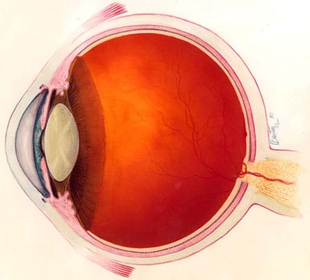
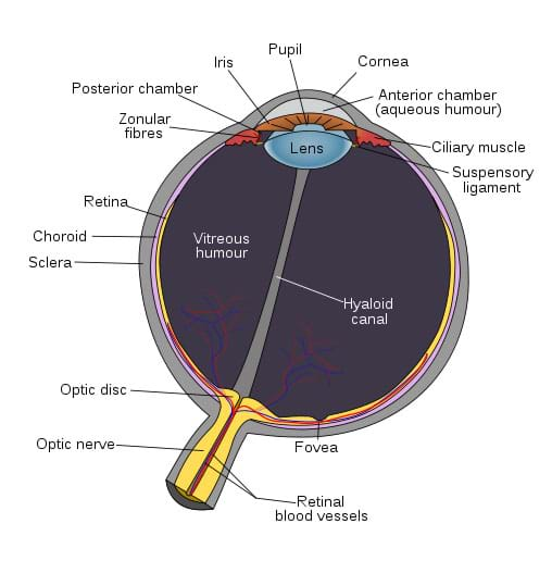
A basic understanding of the parts and functions of the human eye. See Figure 3 and the Vocabulary / Definitions section.
Introduction/Motivation
(Hand out the attached Introduction to the Eyes – Eye-Opening Questions, a set of nine multiple choice questions. Give the students a few minutes to read and circle their answers, before proceeding to introduce the lesson.)
Raise your hand if you have eyes. Okay, that's everyone. Now, how many times a day would you say you use your eyes? A few times? Maybe half the day? Continuously, all day long for almost every task you do? Let's try something. Everyone close their eyes tight, and now shake the hand of the person closest to you. (Let them scramble for a little bit.) Okay, everyone open your eyes and take your seats. I think we all would agree that our eyes are very important for helping us complete even the simplest, everyday tasks. How many of you are wearing glasses or contacts? (Ask some of the students why they are wearing glasses/contacts. Ask if their glasses have ever fallen off or if their contacts have ever fallen out.) Glasses and contacts are great devices because they allow you to see better. But what if you could see perfectly without having to wear them anymore? What if I told you that engineers have developed some biomedical devices that can make that possible? Biomedical engineers design and create devices to help solve some of the common problems with our eyes and our ability to see.

Biomedical engineers develop technologies that help fix eyesight problems, including astigmatism, nearsightedness (myopia) and farsightedness (hyperopic).
- With astigmatism, a person's cornea is not evenly round, which causes light to focus at different distances inside the eye. When looking at an object, some parts may be in focus, while other parts are blurry. To correct this problem, the cornea must be reshaped to be more spherical, so as to correctly focus light on the retina.
- Nearsightedness is the inability to clearly distinguish objects at a distance. This happens when a person's eyeball shape is long, causing light to focus in front of the retina. To correct this, a person must either wear glasses or reshape the cornea to be flatter so that light travels through the lens and focuses correctly on the retina.
- Farsightedness is the inability to clearly distinguish objects up close. This happens when a person's eyeball shape is short (having too flat of a cornea), causing light to focus behind the retina. To correct this, a person must either wear glasses that cause the light to focus sooner or reshape the cornea to be more rounded so that light focuses correctly on the retina.
Engineers have developed many technologies and processes to fix eyesight problems. Some of these technologies can help give a person perfect vision, whether they have trouble seeing far or close, or have trouble focusing. Today, we are going to look in detail at one of these technologies — LASIK surgery. LASIK is a vision correction procedure that uses two different biomedical devices, a microeratome and an excimer laser. The precision surgery reshapes the cornea to change the way light is refracted into the eyeball, so that when it passes through the lens it focuses correctly onto the retina, resulting in clear vision.
For a laser to be used to fix someone's vision, there are some major constraints an biomedical engineer would need to consider and keep in mind while designing. Can you think of any? [Listen to student answers. Possible ideas: something needs to keep the eyeball from twitching, laser needs to be extremely precise, process needs to be specialized for each person's case, needs to be a type of laser that doesn't damage eye tissue.]
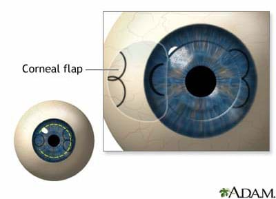
A microkeratome is a very precise, mechanical "shaver" (think of it as a razor for your eye). It has a sharp blade that is guided across the eye, over a set of tracks. It cuts a thin, outer layer of the cornea away from the eye (see Figure 1). The tracks are set on a ring that uses suction to hold the eye still. Once the mid cornea is exposed, it is reshaped by an excimer laser.

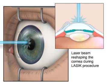
An excimer laser is a "cool" laser because it does not heat up the air around it or the object that it contacts. The laser removes cornea tissue by applying more ultra violet (UV) light than the tissue can tolerate, causing it to break down. The UV light penetrates the cornea less than a nanometer (one billionth of a meter), so only an extremely small amount is removed from the cornea. The laser is also extremely precise. The laser treatment varies for each person, depending on the amount and type of vision correction needed. For nearsightedness, the laser flattens the center of the cornea (see Figure 2); for astigmatism, an oval beam creates a more spherical cornea; and for farsightedness, the laser is applied around the outside of the cornea to make it more rounded.

Engineers have created many other devices for the eyes, as well. Can you think of some devices that you have used before, maybe something to prevent your eyes from being hurt, or to aid them in seeing? (Possible answers: Safety glasses, goggles, sunglasses, binoculars, magnifying glass, microscope, telescope). Refer to the Protect Those Eyes activity to have students design and prototype their own set of protective sports eye wear. Even with perfect vision, some things are still hard or impossible for us to see unless we use devices to aid our vision. Technologies that improve our vision help us to know more about the world around us than we could learn with just our normal eyesight. Vision is an important sense that we need to protect. Let's remember to protect our eyes whenever we are doing an activity that might put them in danger. Even though engineers have designed all kinds of creative devices and equipment to fix our eyes, it's always better to protect them from any injuries first.
(As a class, review the answers to the nine-question introductory handout, providing explanation as provided in the attached Introduction to the Eyes – Answers.)
Lesson Background and Concepts for Teachers
Parts of the Eye
See Figure 3 and the Vocabulary/Definitions section for information on parts of the eye.

More Cool Biomedical Devices for the Eye
To treat uveitis, the inflammation/swelling of the inside of the eye, diabetic macular adema (DME), and age-related macular degeneration (AMD), continuous medical implants are used. When uveitis flares up, it can cause blurred vision, small spots in the field of vision, and cause a person to have difficulty focusing, which can result in the inability to drive a car, go to work or school, or perform other everyday tasks. Almost half of the people who have uveitis develop visual impairment or legal blindness (which accounts for 10 to 15% of all cases of blindness in the US), making it one the leading causes of blindness for middle-aged people in the Western world. (Bausch and Lomb Reisert, 2006) DME is the leading cause of blindness in Americans aged 20-74. It is estimated that 500,000 cases of macular edema exist in the US and ~5,000 new cases develop each year. (SurModics, 2007) AMD affects many people over the age of 65 and almost 50% of people over age 75, with approximately 200,000 new cases each year in the US. (SurModics, 2007)
Retisert is a small medical insert, about the size of a grain of rice, which is surgically implanted in the eye. It is designed to release a precise amount of medicine each day for 2.5 years. It can be put into the eye in less than an hour and the patient can go home the same day. (Bausch and Lomb Reisert, 2006) The benefits of Retisert include continuous and direct medication delivery to the eye to control inflammation with minimal risk to the rest of the body. The negative side is that many patients develop intraocular pressure and cataracts, both of which require further medical treatment (Bausch and Lomb Reisert, 2006).
I-vation is a similar small drug delivery implant. However, it is much different in shape. I-vation is a helical screw made of non-ferrous metal alloy that anchors against the sclera and has a thin cap that rest under the conjunctiva membrane. (SurModics, 2008) I-vation combines biodegradable polymers with drug medications. The chemical release is controlled by the concentration of the drug and the composition of the polymer components. The drug slowly diffuses out of the coating into the eye (SurModics, 2007).
Retinitis pigmentosa is a gene-linked abnormality or degeneration of the rods and cones photoreceptor cells in the eye that can results in loss of night vision in adolescence, peripheral vision as a young adult, and all vision later in life (Source: Hartong, et al., 2006) as well as AMD. While no medical treatment cures this eye condition, future treatments may involve artificial retinas.
Optobioncs Corporation created an artificial retina that can be implanted in the back of the eye to stimulate images in blind patients. This 2 mm-wide chip has 5,000 tiny photodiodes powered by the light that enters the eye. The device creates an electrical signal that stimulates the cells in the back of the eye, which then pass a signal onto the brain. Patients who were completely blind can see blurry shapes; those who could see only blurry shapes can distinguish between teams at a basketball game, and those who had only been able to see their hand in front of their face can make out cars that pass by. This device is still under clinical testing and the long term effects are not fully understood. (McNeely, 2003)
Associated Activities
- Protect Those Eyes - With a specific sport or activity in mind, student teams design and create prototypes for protective eyewear. Students consider possible eye hazards and how engineers incorporate different features and materials into designs to protect the eyes from these risks.
Lesson Closure
Our eyes are an amazing part of our bodies that allows us to interpret so much of the world around us. Our eyes have many intricate parts that work together to give us vision. What are all the parts of the eye? And what are their functions? (Refer to an eye anatomy diagram, if necessary.)
What can go wrong with our eyes? (Listen to student suggestions.) What engineering technologies and inventions can help when a person's vision is impaired? (Possible answers: Prescription glasses and contact lenses, diagnostic/testing equipment and measurement tools, laser surgery, continuous medical implants, drug delivery implants, artificial retinas.)
Engineers develop new biomedical devices that help people with impaired vision to see better. This involves designing within the constraints of the problem: finding the right materials to use, developing tiny functioning parts and tools with the necessary precision, matching the chemical properties of materials with our eyes, and creating machines to build these devices. Engineers are continually redesigning these devices to be more accurate, more effective, and less expensive. The engineering design process is never over, because engineers are always looking for ways to improve the devices we use today. What do you think might be possible 50 years from now?
Vocabulary/Definitions
biomedical engineer: A person who blends traditional engineering techniques with the biological sciences and medicine to improve the quality of human health and life. Biomedical engineers design artificial body parts, medical devices, diagnostic tools, and medical treatment methods.
choroid: A layer of the eye that lines the back of the eye, containing many tiny blood vessels that nourish the retina,
conjunctiva: The thin transparent tissue on the outer surface of the eye that covers the visible part of the eye and lines the eyelids.
constraint: A restriction or limitation.
cornea: The clear, dome-shaped window on the front of the eye. The cornea provides 2/3 of the eye's focusing power. It refracts light towards the lens.
gene: Information about or physical characteristics carried through DNA.
iris: The colored ring that surrounds the pupil. The iris has many tiny muscles that control the size of the pupil, thus regulating the amount of light entering the eye.
LASIK: Acronym for laser-assisted in situ keratomileusis, a procedure that permanently changes the shape of the cornea an excimer laser.
lens: The clear disc behind the iris that helps to focus light, or an image, on the retina (the back of the eye). It is suspended by fibrous strands called zonules.
macula: The small sensitive area of the retina that gives central vision. It is located in the center of the retina and contains the fovea (the center of the macula, giving the sharpest vision).
optic nerve: A bundle of more than one million nerve fibers that carries visual messages from the retina, in the back of the eye, to the brain.
peripheral vision: Seeing things to the sides of the area on which you are focusing. This vision is most sensitive to shapes and movement; objects seen are not in focus.
photodiode: An electrical device that uses light to create an electrical current.
pupil: The black, circular opening in the center of your eye (in the center of the iris) that permits light to enter. The iris adjusts the size of the pupil to control the amount of light that enters the eye. It dilates (becomes larger) in low light to let in more of the available light and constricts (becomes smaller) in bright light.
refract: To change the direction of light passing through.
retina: A thin, light-sensitive tissue lining at the back of the eye that converts light focused on the retina from the lens into electrical impulses that are sent to the brain through the optic nerve.
sclera: The white outer surface of the eye. The sclera is a tough, opaque tissue that protects the inner parts of the eye.
ultraviolet light: Ultraviolet (UV) light travels at a higher frequency than violet light. It is not visible with the human eye. It can cause "sunburn" if exposed for a long period of time.
vitreous: A clear gel that fills the inside of the eyeball. Composed mostly of water, it accounts for about 2/3 of the eye's volume, and gives it shape.
Assessment
Pre-Lesson Assessment
Handout: Have students complete the Introduction to the Eyes – Eye-Opening Questions, a set of nine multiple choice questions, to determine what they currently know or can infer about the eyes. The topics address many features of the eyes as well as interesting facts. Later in the lesson, review the answers as a class, referring to the Introduction to the Eyes – Answers
Post-Introduction Assessment
Biomedical Engineering Benefits Graph: Have all students who wear glasses or contact lenses raise their hands. Also have students who have had eye injuries and gone to the doctor raise their hands. Draw a pie graph on the board with the proportion of students who have been helped by biomedical engineering. To extend this, have all students raise their hands who have a person in their family who wears contact lenses or glasses. (Statistic: In the US, more than half of the population uses some type of lens to correct their vision.)
Lesson Summary Assessment
How LASIK Affects the Eye: As a class, go though each step of the LASIK surgery procedure and ask students which eye parts are being manipulated or affected. Then, have them follow the path of light with the corrected vision into the eye and examine each part that it touches and the function of that part. Use a large diagram of the eye as a visual display (PowerPoint slide, poster or overhead transparency), such as Mark Erickson's annotated drawing of eye anatomy at the Freedom Scientific website: http://www.exquisite-eyes.co.za/common-eye-conditions/eye-anatomy.php.
Lesson Extension Activities
Invite a local LASIK technician to speak to the class about LASIK surgery and eye differences.
Additional Multimedia Support
For a clear, annotated eye anatomy diagram, see Mark Erickson's illustration at the Freedom Scientific website: http://www.freedomscientific.com/resources/vision-anatomy-eye.asp
Show students a one-minute, online animation of the LASIK procedure at the US Food and Drug Administration's LASIK Eye Surgery website: http://www.fda.gov/MedicalDevices/ProductsandMedicalProcedures/SurgeryandLifeSupport/LASIK/ucm061254.htm
Watch a four-minute video of LASIK eye surgery with a doctor explaining each step of procedure as presented by KAMC Channel 28 News in Lubbock, TX. (a bit grainy; choose higher resolution video) at: http://www.westtexaseye.com/products/ImplantableContacs.htm
Alternate six-minute video narrated by doctor (with better video quality) on You Tube, called Dr. Reed Narates Lasik Surgery Video: http://www.youtube.com/watch?v=2m3n7OL09bE
Build your own eye, interactive graphic at Discovery Channel's Human Body Pushing the Limits website: http://dsc.discovery.com/tv/human-body/explorer/explorer.html
A short (2.5 minutes) video from NSF and the University of Michigan on new laser device used to cut corneal flap (to replace microkeratome), developed by engineer, physicist and surgeon, at NSF's News Alert website, called Laser "Scalpel" Improves Popular LASIK Eye Surgery: http://www.nsf.gov/od/lpa/news/press/01/lasik_high.htm
For five excellent drawings of the human eye, one of them interactive, see the National Eye Institute's website called, Diagram of the Eye: http://www.nei.nih.gov/health/eyediagram/index.asp
The National Eye Institute's website for kids provides information and flash activities on parts of the eye (eye diagram, word match, word search), optical illusions, eye safety (safety patrol, protective eyewear, word search) and PDF handouts, called All You Can See: http://isee.nei.nih.gov/
Subscribe
Get the inside scoop on all things Teach Engineering such as new site features, curriculum updates, video releases, and more by signing up for our newsletter!More Curriculum Like This

Students learn about the function and components of the human nervous system, which helps them understand the purpose of our brains, spinal cords, nerves and five senses. In addition, how the nervous system is affected during spaceflight is also discussed.

Students determine their own eyesight and calculate the average eyesight value for the class. They learn about technologies to enhance eyesight and how engineers play an important role in the development of these technologies.

In this activity, students learn about the visual system and then conduct a model experiment to map the visual field response of a Panoptes robot.
References
Anitel, Stefan. "11 Amazing Facts and Myths about Eyes (More Complex than a Photo Camera)" Published December 27, 2007. Softpedia. Accessed February 11, 2009. http://news.softpedia.com/news/10-Amazing-Facts-and-Myths-About-Eyes-74813.shtml
Bausch and Lomb. Retisert (fluocinolone acetonide intravitreal implant) Patient Section Home. 2006. Accessed February 11, 2009. http://www.retisert.com/patient_home.html
Diagram of the Eye. Last modified October 2008. National Eye Institute, National Institutes of Health. Accessed February 11, 2009. (source of some vocabulary definitions, with modifications) http://www.nei.nih.gov/health/eyediagram/index.asp
Eye Anatomy: Parts and Functions. St. Luke's Cataract and Laser Institute. Accessed November 3, 2010. http://www.stlukeseye.com/anatomy/
Hartong, D.T., Berson, E.L. and Dyria, T.P. Retinitis Pigmentosa. Published November 18, 2006. Lancet 368 (9549); 1795-809. PubMed.gov, NCBI, NLM, National Institutes of Health. Accessed February 11, 2009. http://www.ncbi.nlm.nih.gov/pubmed/17113430
Learning about LASIK. Updated September 18, 2008. US Food and Drug Administration and Center for Devices and Radiological Health, US Department of Health and Human Services. Accessed February 11, 2009. http://www.fda.gov/
McNeely, Gretchen. "Optobionics stays focused as retina implant process plods along." Published June 16, 2003. Small Times. Accessed February 11, 2009. http://www.smalltimes.com/Articles/Article_Display.cfm?ARTICLE_ID=268807&p=109
Tyson, Jeff. How LASIK Works. September 19, 2001. HowStuffWorks.com. Accessed February 11, 2009. http://health.howstuffworks.com/lasik.htm
Understanding LASIK. The Vision Correction Website. Internet Media Service Inc. Accessed February 11, 2009. (good description and diagrams of surgery steps, plus link to video) http://www.lasersite.com/lasik/index.htm
Copyright
© 2008 by Regents of the University of ColoradoContributors
William Surles; Lesley Herrmann; Malinda Schaefer Zarske; Denise W. CarlsonSupporting Program
Integrated Teaching and Learning Program, College of Engineering, University of Colorado BoulderAcknowledgements
The contents of this digital library curriculum were developed under a grant from the Fund for the Improvement of Postsecondary Education (FIPSE), U.S. Department of Education and National Science Foundation GK-12 grant no. 0338326. However, these contents do not necessarily represent the policies of the Department of Education or National Science Foundation, and you should not assume endorsement by the federal government.
Last modified: May 14, 2022


User Comments & Tips